1ORW | pdb_00001orw

Crystal Structure of Porcine Dipeptidyl Peptidase IV (CD26) in Complex with a Peptidomimetic Inhibitor


Experimental Data Snapshot

  • Method: X-RAY DIFFRACTION
  • Resolution: 2.84 Å
  • R-Value Free: 
    0.246 (Depositor), 0.235 (DCC) 
  • R-Value Work: 
    0.187 (Depositor), 0.177 (DCC) 
  • R-Value Observed: 
    0.187 (Depositor) 

wwPDB Validation 3D Report Full Report

Validation slider image for 1ORW

Ligand Structure Quality Assessment 


This is version 2.1 of the entry. See complete history

Literature

The Crystal Structure of Dipeptidyl Peptidase IV (CD26) Reveals its Functional Regulation and Enzymatic Mechanism

Engel, M.Hoffmann, T.Wagner, L.Wermann, M.Heiser, U.Kiefersauer, R.Huber, R.Bode, W.Demuth, H.U.Brandstetter, H.

(2003) Proc Natl Acad Sci U S A 100: 5063-5068

  • DOI: https://doi.org/10.1073/pnas.0230620100
  • Primary Citation Related Structures: 
    1ORV, 1ORW

  • PubMed Abstract: 

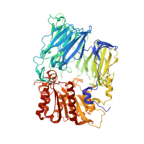
    The membrane-bound glycoprotein dipeptidyl peptidase IV (DP IV, CD26) is a unique multifunctional protein, acting as receptor, binding and proteolytic molecule. We have determined the sequence and 1.8 A crystal structure of native DP IV prepared from porcine kidney. The crystal structure reveals a 2-2-2 symmetric tetrameric assembly which depends on the natively glycosylated beta-propeller blade IV. The crystal structure indicates that tetramerization of DP IV is a key mechanism to regulate its interaction with other components. Each subunit comprises two structural domains, the N-terminal eight-bladed beta-propeller with open Velcro topology and the C-terminal alpha/beta-hydrolase domain. Analogy with the structurally related POP and tricorn protease suggests that substrates access the buried active site through the beta-propeller tunnel while products leave the active site through a separate side exit. A dipeptide mimicking inhibitor complexed to the active site discloses key determinants for substrate recognition, including a Glu-Glu motif that distinguishes DP IV as an aminopeptidase and an oxyanion trap that binds and activates the P(2)-carbonyl oxygen necessary for efficient postproline cleavage. We discuss active and nonactive site-directed inhibition strategies of this pharmaceutical target protein.


  • Organizational Affiliation
    • Max-Planck-Institut für Biochemie, Abt. Strukturforschung, D-82152 Martinsried, Germany.

Macromolecule Content 

  • Total Structure Weight: 347.13 kDa 
  • Atom Count: 25,262 
  • Modeled Residue Count: 2,912 
  • Deposited Residue Count: 2,912 
  • Unique protein chains: 1

Macromolecules

Find similar proteins by:|  3D Structure
Entity ID: 1
MoleculeChains  Sequence LengthOrganismDetailsImage
dipeptidyl peptidase IV
A, B, C, D
728Sus scrofaMutation(s): 0 
EC: 3.4.14.5
UniProt
Find proteins for P22411 (Sus scrofa)
Explore P22411 
Go to UniProtKB:  P22411
Entity Groups
Sequence Clusters30% Identity50% Identity70% Identity90% Identity95% Identity100% Identity
UniProt GroupP22411
Glycosylation
Glycosylation Sites: 6
Sequence Annotations
Expand
Reference Sequence

Oligosaccharides

Help  
Entity ID: 2
MoleculeChains Length2D Diagram GlycosylationD Interactions
2-acetamido-2-deoxy-beta-D-glucopyranose-(1-4)-2-acetamido-2-deoxy-beta-D-glucopyranose
E, F, H, I, K
E, F, H, I, K, L, M
2N-Glycosylation
Glycosylation Resources
GlyTouCan: G42666HT
GlyCosmos: G42666HT
GlyGen: G42666HT
Entity ID: 3
MoleculeChains Length2D Diagram GlycosylationD Interactions
beta-D-mannopyranose-(1-4)-2-acetamido-2-deoxy-beta-D-glucopyranose-(1-4)-2-acetamido-2-deoxy-beta-D-glucopyranose
G, J
3N-Glycosylation
Glycosylation Resources
GlyTouCan: G15407YE
GlyCosmos: G15407YE
GlyGen: G15407YE

Small Molecules

Ligands 4 Unique
IDChains Name / Formula / InChI Key2D Diagram3D Interactions
PHI

Query on PHI


Download:Ideal Coordinates CCD File
HA [auth C],
MA [auth D],
S [auth A],
Z [auth B]
IODO-PHENYLALANINE
C9 H10 I N O2
PZNQZSRPDOEBMS-QMMMGPOBSA-N
NAG

Query on NAG


Download:Ideal Coordinates CCD File
BA [auth C]
CA [auth C]
DA [auth C]
EA [auth C]
FA [auth C]
BA [auth C],
CA [auth C],
DA [auth C],
EA [auth C],
FA [auth C],
JA [auth D],
KA [auth D],
N [auth A],
O [auth A],
P [auth A],
Q [auth A],
U [auth B],
V [auth B],
W [auth B],
X [auth B]
2-acetamido-2-deoxy-beta-D-glucopyranose
C8 H15 N O6
OVRNDRQMDRJTHS-FMDGEEDCSA-N
P2Y

Query on P2Y


Download:Ideal Coordinates CCD File
AA [auth B],
IA [auth C],
NA [auth D],
T [auth A]
(2S)-PYRROLIDIN-2-YLMETHYLAMINE
C5 H12 N2
AUKXFNABVHIUAC-YFKPBYRVSA-N
SO4

Query on SO4


Download:Ideal Coordinates CCD File
GA [auth C],
LA [auth D],
R [auth A],
Y [auth B]
SULFATE ION
O4 S
QAOWNCQODCNURD-UHFFFAOYSA-L

Experimental Data & Validation

Experimental Data

  • Method: X-RAY DIFFRACTION
  • Resolution: 2.84 Å
  • R-Value Free:  0.246 (Depositor), 0.235 (DCC) 
  • R-Value Work:  0.187 (Depositor), 0.177 (DCC) 
  • R-Value Observed: 0.187 (Depositor) 
Space Group: P 1
Unit Cell:
Length ( Å )Angle ( ˚ )
a = 61.97α = 112.65
b = 117.71β = 94.81
c = 133.56γ = 91.17
Software Package:
Software NamePurpose
CNSrefinement
DENZOdata reduction
SCALEPACKdata scaling
MAINphasing

Structure Validation

View Full Validation Report


Ligand Structure Quality Assessment 


Entry History 

Deposition Data

Revision History  (Full details and data files)

  • Version 1.0: 2003-05-06
    Type: Initial release
  • Version 1.1: 2008-04-29
    Changes: Version format compliance
  • Version 1.2: 2011-07-13
    Changes: Non-polymer description, Version format compliance
  • Version 2.0: 2020-07-29
    Type: Remediation
    Reason: Carbohydrate remediation
    Changes: Advisory, Atomic model, Data collection, Derived calculations, Structure summary
  • Version 2.1: 2025-03-26
    Changes: Advisory, Data collection, Database references, Structure summary