Structure of the heterodimeric neurotoxic complex viperotoxin F (RV-4/RV-7) from the venom of Vipera russelli formosensis at 1.9 A resolution.
Perbandt, M., Tsai, I.H., Fuchs, A., Banumathi, S., Rajashankar, K.R., Georgieva, D., Kalkura, N., Singh, T.P., Genov, N., Betzel, C.(2003) Acta Crystallogr D Biol Crystallogr 59: 1679-1687
- PubMed: 14501106 Search on PubMed
- DOI: https://doi.org/10.1107/s0907444903014987
- Primary Citation Related Structures:
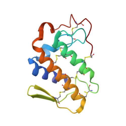
1OQS - PubMed Abstract:
The presynaptic viperotoxin F is the major lethal component of the venom of Vipera russelli formosensis (Taiwan viper). It is a heterodimer of two highly homologous (65% identity) but oppositely charged subunits: a basic and neurotoxic PLA(2) (RV-4) and an acidic non-toxic component with a very low enzymatic activity (RV-7). The crystal structure of the complex has been determined by molecular replacement and refined to 1.9 A resolution and an R factor of 22.3% with four RV-4/RV-7 complexes in the asymmetric unit, which do not exhibit any local point-group symmetry. The complex formation decreases the accessible surface area of the two subunits by approximately 1425 A(2). Both PLA(2)s are predicted to have very low, if any, anticoagulant activity. The structure of viperotoxin F is compared with that of the heterodimeric neurotoxin vipoxin from the venom of another viper, V. ammodytes meridionalis. The structural basis for the differences between the pharmacological activities of the two toxins is discussed. The neutralization of the negative charge of the major ligand for Ca(2+), Asp49, by intersubunit salt bridges is probably a common mechanism of self-stabilization of heterodimeric Viperinae snake-venom neurotoxins in the absence of bound calcium.
- Institute of Biochemistry and Molecular Biology I, University Hospital Hamburg-Eppendorf, c/o DESY, Building 22a, Notkestrasse 85, 22603 Hamburg, Germany.
Organizational Affiliation: