1OKY | pdb_00001oky

Structure of human PDK1 kinase domain in complex with staurosporine


Experimental Data Snapshot

  • Method: X-RAY DIFFRACTION
  • Resolution: 2.30 Å
  • R-Value Free: 
    0.255 (Depositor), 0.240 (DCC) 
  • R-Value Work: 
    0.219 (Depositor), 0.207 (DCC) 
  • R-Value Observed: 
    0.219 (Depositor) 

Starting Model: experimental
View more details

wwPDB Validation 3D Report Full Report

Validation slider image for 1OKY

Ligand Structure Quality Assessment 


This is version 1.5 of the entry. See complete history

Literature

Structural Basis for Ucn-01 (7-Hydroxystaurosporine) Specificity and Pdk1 (3-Phosphoinositide-Dependent Protein Kinase-1) Inhibition

Komander, D.Kular, G.S.Bain, J.Elliot, M.Alessi, D.R.Van Aalten, D.M.F.

(2003) Biochem J 375: 255

  • DOI: https://doi.org/10.1042/BJ20031119
  • Primary Citation Related Structures: 
    1OKY, 1OKZ

  • PubMed Abstract: 

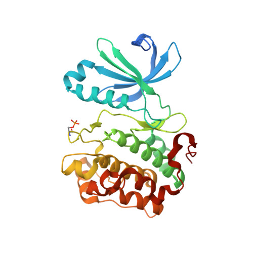
    PDK1 (3-phosphoinositide-dependent protein kinase-1) is a member of the AGC (cAMP-dependent, cGMP-dependent, protein kinase C) family of protein kinases, and has a key role in insulin and growth-factor signalling through phosphorylation and subsequent activation of a number of other AGC kinase family members, such as protein kinase B. The staurosporine derivative UCN-01 (7-hydroxystaurosporine) has been reported to be a potent inhibitor for PDK1, and is currently undergoing clinical trials for the treatment of cancer. Here, we report the crystal structures of staurosporine and UCN-01 in complex with the kinase domain of PDK1. We show that, although staurosporine and UCN-01 interact with the PDK1 active site in an overall similar manner, the UCN-01 7-hydroxy group, which is not present in staurosporine, generates direct and water-mediated hydrogen bonds with active-site residues. Inhibition data from UCN-01 tested against a panel of 29 different kinases show a different pattern of inhibition compared with staurosporine. We discuss how these differences in inhibition could be attributed to specific interactions with the additional 7-hydroxy group, as well as the size of the 7-hydroxy-group-binding pocket. This information could lead to opportunities for structure-based optimization of PDK1 inhibitors.


  • Organizational Affiliation
    • Division of Biological Chemistry and Molecular Microbiology, School of Life Sciences, University of Dundee, Dundee DD1 5EH, Scotland, UK.

Macromolecule Content 

  • Total Structure Weight: 36.72 kDa 
  • Atom Count: 2,475 
  • Modeled Residue Count: 283 
  • Deposited Residue Count: 310 
  • Unique protein chains: 1

Macromolecules

Find similar proteins by:|  3D Structure
Entity ID: 1
MoleculeChains  Sequence LengthOrganismDetailsImage
3-PHOSPHOINOSITIDE DEPENDENT PROTEIN KINASE 1310Homo sapiensMutation(s): 0 
EC: 2.7.1.37 (PDB Primary Data), 2.7.11.1 (UniProt)
UniProt & NIH Common Fund Data Resources
Find proteins for O15530 (Homo sapiens)
Explore O15530 
Go to UniProtKB:  O15530
PHAROS:  O15530
GTEx:  ENSG00000140992 
Entity Groups
Sequence Clusters30% Identity50% Identity70% Identity90% Identity95% Identity100% Identity
UniProt GroupO15530
Sequence Annotations
Expand
Reference Sequence

Small Molecules

Ligands 3 Unique
IDChains Name / Formula / InChI Key2D Diagram3D Interactions
STU

Query on STU


Download:Ideal Coordinates CCD File
J [auth A]STAUROSPORINE
C28 H26 N4 O3
HKSZLNNOFSGOKW-FYTWVXJKSA-N
SO4

Query on SO4


Download:Ideal Coordinates CCD File
E [auth A],
F [auth A],
G [auth A],
H [auth A],
I [auth A]
SULFATE ION
O4 S
QAOWNCQODCNURD-UHFFFAOYSA-L
GOL

Query on GOL


Download:Ideal Coordinates CCD File
B [auth A],
C [auth A],
D [auth A]
GLYCEROL
C3 H8 O3
PEDCQBHIVMGVHV-UHFFFAOYSA-N
Modified Residues  1 Unique
IDChains TypeFormula2D DiagramParent
SEP
Query on SEP
A
L-PEPTIDE LINKINGC3 H8 N O6 PSER
Binding Affinity Annotations 
IDSourceBinding Affinity
STU BindingDB:  1OKY IC50: 5.6 (nM) from 1 assay(s)

Experimental Data & Validation

Experimental Data

  • Method: X-RAY DIFFRACTION
  • Resolution: 2.30 Å
  • R-Value Free:  0.255 (Depositor), 0.240 (DCC) 
  • R-Value Work:  0.219 (Depositor), 0.207 (DCC) 
  • R-Value Observed: 0.219 (Depositor) 
Space Group: P 32 2 1
Unit Cell:
Length ( Å )Angle ( ˚ )
a = 124.172α = 90
b = 124.172β = 90
c = 47.314γ = 120
Software Package:
Software NamePurpose
CNSrefinement
DENZOdata reduction
SCALEPACKdata scaling
AMoREphasing

Structure Validation

View Full Validation Report


Ligand Structure Quality Assessment 


Entry History 

Deposition Data

Revision History  (Full details and data files)

  • Version 1.0: 2004-07-29
    Type: Initial release
  • Version 1.1: 2011-05-08
    Changes: Version format compliance
  • Version 1.2: 2011-07-13
    Changes: Version format compliance
  • Version 1.3: 2019-04-24
    Changes: Advisory, Data collection, Derived calculations, Other, Source and taxonomy
  • Version 1.4: 2023-12-13
    Changes: Advisory, Data collection, Database references, Other, Refinement description
  • Version 1.5: 2024-10-23
    Changes: Structure summary