1OGL

The crystal structure of native Trypanosoma cruzi dUTPase


Experimental Data Snapshot

  • Method: X-RAY DIFFRACTION
  • Resolution: 2.40 Å
  • R-Value Free: 0.263 
  • R-Value Work: 0.203 
  • R-Value Observed: 0.206 

wwPDB Validation   3D Report Full Report


This is version 1.2 of the entry. See complete history


Literature

The Crystal Structure of Trypanosoma Cruzi Dutpase Reveals a Novel Dutp/Dudp Binding Fold

Harkiolaki, M.Dodson, E.J.Bernier-Villamor, V.Turkenburg, J.P.Gonzalez-Pacanowska, D.Wilson, K.S.

(2004) Structure 12: 41

  • DOI: https://doi.org/10.1016/j.str.2003.11.016
  • Primary Citation of Related Structures:  
    1OGK, 1OGL

  • PubMed Abstract: 

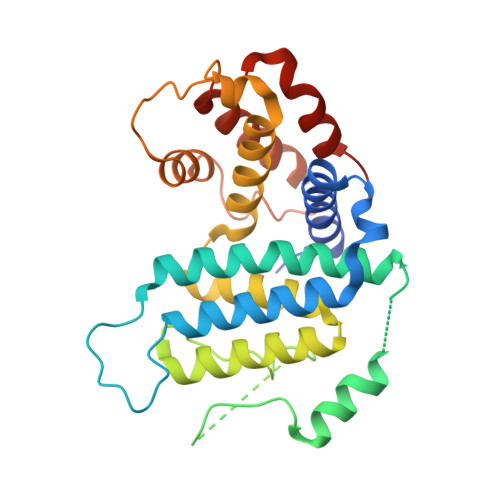
    dUTPase is an essential enzyme involved with nucleotide metabolism and replication. We report here the X-ray structure of Trypanosoma cruzi dUTPase in its native conformation and as a complex with dUDP. These reveal a novel protein fold that displays no structural similarities to previously described dUTPases. The molecular unit is a dimer with two active sites. Nucleotide binding promotes extensive structural rearrangements, secondary structure remodeling, and rigid body displacements of 20 A or more, which effectively bury the substrate within the enzyme core for the purpose of hydrolysis. The molecular complex is a trapped enzyme-substrate arrangement which clearly demonstrates structure-induced specificity and catalytic potential. This enzyme is a novel dUTPase and therefore a potential drug target in the treatment of Chagas' disease.


  • Organizational Affiliation

    Structural Biology Laboratory, Department of Chemistry, University of York, Heslington, York YO10 5YW, UK.


Macromolecules
Find similar proteins by:  (by identity cutoff)  |  3D Structure
Entity ID: 1
MoleculeChains Sequence LengthOrganismDetailsImage
DEOXYURIDINE TRIPHOSPHATASE283Trypanosoma cruziMutation(s): 0 
EC: 3.6.1.23
UniProt
Find proteins for O15923 (Trypanosoma cruzi)
Explore O15923 
Go to UniProtKB:  O15923
Entity Groups  
Sequence Clusters30% Identity50% Identity70% Identity90% Identity95% Identity100% Identity
UniProt GroupO15923
Sequence Annotations
Expand
  • Reference Sequence
Experimental Data & Validation

Experimental Data

  • Method: X-RAY DIFFRACTION
  • Resolution: 2.40 Å
  • R-Value Free: 0.263 
  • R-Value Work: 0.203 
  • R-Value Observed: 0.206 
  • Space Group: P 63 2 2
Unit Cell:
Length ( Å )Angle ( ˚ )
a = 136.434α = 90
b = 136.434β = 90
c = 68.705γ = 120
Software Package:
Software NamePurpose
REFMACrefinement
DENZOdata reduction
SCALEPACKdata scaling
SOLVEphasing
MLPHAREphasing
AMoREphasing

Structure Validation

View Full Validation Report


Entry History 

Deposition Data

Revision History  (Full details and data files)

  • Version 1.0: 2004-01-22
    Type: Initial release
  • Version 1.1: 2011-05-08
    Changes: Version format compliance
  • Version 1.2: 2011-07-13
    Changes: Version format compliance