1OGF

The Structure of Bacillus subtilis RbsD complexed with glycerol


Experimental Data Snapshot

  • Method: X-RAY DIFFRACTION
  • Resolution: 2.30 Å
  • R-Value Free: 0.250 
  • R-Value Work: 0.215 
  • R-Value Observed: 0.215 

wwPDB Validation   3D Report Full Report


This is version 1.3 of the entry. See complete history


Literature

Crystal Structures of Rbsd Leading to the Identification of Cytoplasmic Sugar-Binding Proteins with a Novel Folding Architecture

Kim, M.-S.Shin, J.Lee, W.Lee, H.-S.Oh, B.-H.

(2003) J Biol Chem 278: 28173

  • DOI: https://doi.org/10.1074/jbc.M304523200
  • Primary Citation of Related Structures:  
    1OGC, 1OGD, 1OGE, 1OGF

  • PubMed Abstract: 

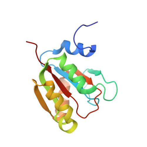
    RbsD is the only protein whose biochemical function is unknown among the six gene products of the rbs operon involved in the active transport of ribose. FucU, a paralogue of RbsD conserved from bacteria to human, is also the only protein whose function is unknown among the seven gene products of the l-fucose regulon. Here we report the crystal structures of Bacillus subtilis RbsD, which reveals a novel decameric toroidal assembly of the protein. Nuclear magnetic resonance and other studies on RbsD reveal that the intersubunit cleft of the protein binds specific forms of d-ribose, but it does not have an enzyme activity toward the sugar. Likewise, FucU binds l-fucose but lacks an enzyme activity toward this sugar. We conclude that RbsD and FucU are cytoplasmic sugar-binding proteins, a novel class of proteins whose functional role may lie in helping influx of the sugar substrates.


  • Organizational Affiliation

    Center for Biomolecular Recognition and Division of Molecular and Life Science, Department of Life Science, Pohang University of Science and Technology, Pohang, Kyungbuk, 790-784, Korea.


Macromolecules
Find similar proteins by:  (by identity cutoff)  |  3D Structure
Entity ID: 1
MoleculeChains Sequence LengthOrganismDetailsImage
HIGH AFFINITY RIBOSE TRANSPORT PROTEIN RBSD
A, B, C, D, E
131Bacillus subtilisMutation(s): 0 
UniProt
Find proteins for P36946 (Bacillus subtilis (strain 168))
Explore P36946 
Go to UniProtKB:  P36946
Entity Groups  
Sequence Clusters30% Identity50% Identity70% Identity90% Identity95% Identity100% Identity
UniProt GroupP36946
Sequence Annotations
Expand
  • Reference Sequence
Experimental Data & Validation

Experimental Data

  • Method: X-RAY DIFFRACTION
  • Resolution: 2.30 Å
  • R-Value Free: 0.250 
  • R-Value Work: 0.215 
  • R-Value Observed: 0.215 
  • Space Group: C 1 2 1
Unit Cell:
Length ( Å )Angle ( ˚ )
a = 122.314α = 90
b = 106.222β = 128.5
c = 82.648γ = 90
Software Package:
Software NamePurpose
CNSrefinement

Structure Validation

View Full Validation Report


Entry History 

Deposition Data

Revision History  (Full details and data files)

  • Version 1.0: 2003-09-01
    Type: Initial release
  • Version 1.1: 2011-05-08
    Changes: Version format compliance
  • Version 1.2: 2011-07-13
    Changes: Version format compliance
  • Version 1.3: 2019-05-22
    Changes: Data collection, Other, Refinement description