1OFI | pdb_00001ofi

Asymmetric complex between HslV and I-domain deleted HslU (H. influenzae)


Experimental Data Snapshot

  • Method: X-RAY DIFFRACTION
  • Resolution: 3.20 Å
  • R-Value Free: 
    0.327 (Depositor) 
  • R-Value Work: 
    0.232 (Depositor) 
  • R-Value Observed: 
    0.232 (Depositor) 

Starting Model: experimental
View more details

wwPDB Validation 3D Report Full Report

Validation slider image for 1OFI

This is version 1.4 of the entry. See complete history

Literature

Structure and Reactivity of an Asymmetric Complex between Hslv and I-Domain Deleted Hslu, a Prokaryotic Homolog of the Eukaryotic Proteasome

Kwon, A.R.Kessler, B.M.Overkleeft, H.S.Mckay, D.B.

(2003) J Mol Biology 330: 185

  • DOI: https://doi.org/10.1016/s0022-2836(03)00580-1
  • Primary Citation Related Structures: 
    1OFH, 1OFI

  • PubMed Abstract: 

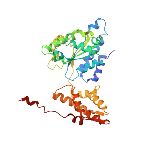
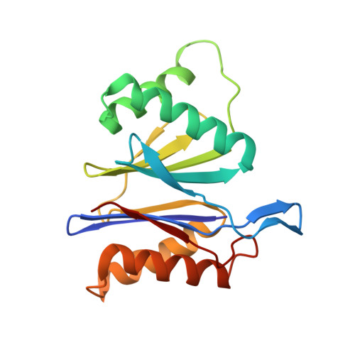
    In the prokaryotic homolog of the eukaryotic proteasome, HslUV, the "double donut" HslV protease is allosterically activated by HslU, an AAA protein of the Clp/Hsp100 family consisting of three (amino-terminal, carboxy-terminal, and intermediate) domains. The intermediate domains of HslU, which extend like tentacles from the hexameric ring formed by the amino-terminal and carboxy-terminal domains, have been deleted; an asymmetric HslU(DeltaI)(6)HslV(12) complex has been crystallized; and the structure has been solved to 2.5A resolution, revealing an assembly in which a HslU(DeltaI) hexamer binds one end of the HslV dodecamer. The conformation of the protomers of the HslU(DeltaI)-complexed HslV hexamer is similar to that in the symmetric wild-type HslUV complex, while the protomer conformation of the uncomplexed HslV hexamer is similar to that of HslV alone. Reaction in the crystals with a vinyl sulfone inhibitor reveals that the HslU(DeltaI)-complexed HslV hexamer is active, while the uncomplexed HslV hexamer is inactive. These results confirm that HslV can be activated by binding of a hexameric HslU(DeltaI)(6) ring lacking the I domains, that activation is effected through a conformational change in HslV rather than through alteration of the size of the entry channel into the protease catalytic cavity, and that the two HslV(6) rings in the protease dodecamer are activated independently rather than cooperatively.


  • Organizational Affiliation
    • Department of Structural Biology, Stanford University School of Medicine, Sherman Fairchild Building, Stanford, CA 94305, USA.

Macromolecule Content 

  • Total Structure Weight: 219.83 kDa 
  • Atom Count: 15,050 
  • Modeled Residue Count: 1,934 
  • Deposited Residue Count: 1,974 
  • Unique protein chains: 2

Macromolecules

Find similar proteins by:|  3D Structure
Entity ID: 1
MoleculeChains  Sequence LengthOrganismDetailsImage
ATP-DEPENDENT HSL PROTEASE ATP-BINDING SUBUNIT HSLU
A, B, C
310Haemophilus influenzae Rd KW20Mutation(s): 0 
UniProt
Find proteins for P43773 (Haemophilus influenzae (strain ATCC 51907 / DSM 11121 / KW20 / Rd))
Explore P43773 
Go to UniProtKB:  P43773
Entity Groups
Sequence Clusters30% Identity50% Identity70% Identity90% Identity95% Identity100% Identity
UniProt GroupP43773
Sequence Annotations
Expand
Reference Sequence
Find similar proteins by:|  3D Structure
Entity ID: 2
MoleculeChains  Sequence LengthOrganismDetailsImage
ATP-DEPENDENT PROTEASE HSLV174Haemophilus influenzae Rd KW20Mutation(s): 0 
EC: 3.4.25 (PDB Primary Data), 3.4.25.2 (UniProt)
UniProt
Find proteins for P43772 (Haemophilus influenzae (strain ATCC 51907 / DSM 11121 / KW20 / Rd))
Explore P43772 
Go to UniProtKB:  P43772
Entity Groups
Sequence Clusters30% Identity50% Identity70% Identity90% Identity95% Identity100% Identity
UniProt GroupP43772
Sequence Annotations
Expand
Reference Sequence

Small Molecules

Ligands 4 Unique
IDChains Name / Formula / InChI Key2D Diagram3D Interactions
LVS

Query on LVS


Download:Ideal Coordinates CCD File
V [auth G],
X [auth H],
Z [auth I]
4-IODO-3-NITROPHENYL ACETYL-LEUCINYL-LEUCINYL-LEUCINYL-VINYLSULFONE
C28 H43 I N4 O8 S
RSYTXXARKSZDKY-PUHATCMVSA-N
ADP

Query on ADP


Download:Ideal Coordinates CCD File
J [auth A],
N [auth B],
R [auth C]
ADENOSINE-5'-DIPHOSPHATE
C10 H15 N5 O10 P2
XTWYTFMLZFPYCI-KQYNXXCUSA-N
PO4

Query on PO4


Download:Ideal Coordinates CCD File
L [auth A],
P [auth B],
T [auth C]
PHOSPHATE ION
O4 P
NBIIXXVUZAFLBC-UHFFFAOYSA-K
MG

Query on MG


Download:Ideal Coordinates CCD File
AA [auth I]
BA [auth L]
CA [auth M]
DA [auth N]
K [auth A]
AA [auth I],
BA [auth L],
CA [auth M],
DA [auth N],
K [auth A],
M [auth A],
O [auth B],
Q [auth B],
S [auth C],
U [auth C],
W [auth G],
Y [auth H]
MAGNESIUM ION
Mg
JLVVSXFLKOJNIY-UHFFFAOYSA-N

Experimental Data & Validation

Experimental Data

  • Method: X-RAY DIFFRACTION
  • Resolution: 3.20 Å
  • R-Value Free:  0.327 (Depositor) 
  • R-Value Work:  0.232 (Depositor) 
  • R-Value Observed: 0.232 (Depositor) 
Space Group: P 6
Unit Cell:
Length ( Å )Angle ( ˚ )
a = 190.177α = 90
b = 190.177β = 90
c = 114.511γ = 120
Software Package:
Software NamePurpose
CNSrefinement
DENZOdata reduction
SCALEPACKdata scaling
AMoREphasing

Structure Validation

View Full Validation Report


Entry History 

Deposition Data

Revision History  (Full details and data files)

  • Version 1.0: 2003-07-03
    Type: Initial release
  • Version 1.1: 2011-05-08
    Changes: Version format compliance
  • Version 1.2: 2011-07-13
    Changes: Version format compliance
  • Version 1.3: 2023-12-13
    Changes: Data collection, Database references, Derived calculations, Refinement description
  • Version 1.4: 2024-11-20
    Changes: Structure summary