1O99 | pdb_00001o99

CRYSTAL STRUCTURE OF THE S62A MUTANT OF PHOSPHOGLYCERATE MUTASE FROM BACILLUS STEAROTHERMOPHILUS COMPLEXED WITH 2-PHOSPHOGLYCERATE


Experimental Data Snapshot

  • Method: X-RAY DIFFRACTION
  • Resolution: 2.65 Å
  • R-Value Free: 
    0.242 (Depositor), 0.229 (DCC) 
  • R-Value Work: 
    0.199 (Depositor), 0.187 (DCC) 
  • R-Value Observed: 
    0.199 (Depositor) 

Starting Model: experimental
View more details

wwPDB Validation 3D Report Full Report

Validation slider image for 1O99

Ligand Structure Quality Assessment 


This is version 1.3 of the entry. See complete history

Literature

Insights Into the Catalytic Mechanism of Cofactor-Independent Phosphoglycerate Mutase from X-Ray Crystallography, Simulated Dynamics and Molecular Modeling

Rigden, D.J.Mello, L.V.Lamani, E.Littlejohn, J.E.Jedrzejas, M.J.

(2003) J Mol Biology 328: 909

  • DOI: https://doi.org/10.1016/s0022-2836(03)00350-4
  • Primary Citation Related Structures: 
    1O98, 1O99

  • PubMed Abstract: 

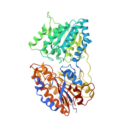
    Phosphoglycerate mutases catalyze the isomerization of 2 and 3-phosphoglycerates, and are essential for glucose metabolism in most organisms. Here, we further characterize the 2,3-bisphosphoglycerate-independent phosphoglycerate mutase (iPGM) from Bacillus stearothermophilus by determination of a high-resolution (1.4A) crystal structure of the wild-type enzyme and the crystal structure of its S62A mutant. The mutant structure surprisingly showed the replacement of one of the two catalytically essential manganese ions with a water molecule, offering an additional possible explanation for its lack of catalytic activity. Crystal structures invariably show substrate phosphoglycerate to be entirely buried in a deep cleft between the two iPGM domains. Flexibility analyses were therefore employed to reveal the likely route of substrate access to the catalytic site through an aperture created in the enzyme's surface during certain stages of the catalytic process. Several conserved residues lining this aperture may contribute to orientation of the substrate as it enters. Factors responsible for the retention of glycerate within the phosphoenzyme structure in the proposed mechanism are identified by molecular modeling of the glycerate complex of the phosphoenzyme. Taken together, these results allow for a better understanding of the mechanism of action of iPGMs. Many of the results are relevant to a series of evolutionarily related enzymes. These studies will facilitate the development of iPGM inhibitors which, due to the demonstrated importance of this enzyme in many bacteria, would be of great potential clinical significance.


  • Organizational Affiliation
    • National Centre of Genetic Resources and Biotechnology, Cenargen/Embrapa, S.A.I.N. Parque Rural, Final W5, Asa Norte, 70770-900 Brasília, Brazil.

Macromolecule Content 

  • Total Structure Weight: 57.82 kDa 
  • Atom Count: 4,380 
  • Modeled Residue Count: 510 
  • Deposited Residue Count: 511 
  • Unique protein chains: 1

Macromolecules

Find similar proteins by:|  3D Structure
Entity ID: 1
MoleculeChains  Sequence LengthOrganismDetailsImage
2,3-BISPHOSPHOGLYCERATE-INDEPENDENT PHOSPHOGLYCERATE MUTASE511Geobacillus stearothermophilusMutation(s): 1 
EC: 5.4.2.1 (PDB Primary Data), 5.4.2.12 (UniProt)
UniProt
Find proteins for Q9X519 (Geobacillus stearothermophilus)
Explore Q9X519 
Go to UniProtKB:  Q9X519
Entity Groups
Sequence Clusters30% Identity50% Identity70% Identity90% Identity95% Identity100% Identity
UniProt GroupQ9X519
Sequence Annotations
Expand
Reference Sequence

Small Molecules

Ligands 3 Unique
IDChains Name / Formula / InChI Key2D Diagram3D Interactions
2PG

Query on 2PG


Download:Ideal Coordinates CCD File
C [auth A]2-PHOSPHOGLYCERIC ACID
C3 H7 O7 P
GXIURPTVHJPJLF-UWTATZPHSA-N
SO4

Query on SO4


Download:Ideal Coordinates CCD File
D [auth A]
E [auth A]
F [auth A]
G [auth A]
H [auth A]
D [auth A],
E [auth A],
F [auth A],
G [auth A],
H [auth A],
I [auth A]
SULFATE ION
O4 S
QAOWNCQODCNURD-UHFFFAOYSA-L
MN

Query on MN


Download:Ideal Coordinates CCD File
B [auth A]MANGANESE (II) ION
Mn
WAEMQWOKJMHJLA-UHFFFAOYSA-N

Experimental Data & Validation

Experimental Data

  • Method: X-RAY DIFFRACTION
  • Resolution: 2.65 Å
  • R-Value Free:  0.242 (Depositor), 0.229 (DCC) 
  • R-Value Work:  0.199 (Depositor), 0.187 (DCC) 
  • R-Value Observed: 0.199 (Depositor) 
Space Group: C 2 2 21
Unit Cell:
Length ( Å )Angle ( ˚ )
a = 58.712α = 90
b = 207.758β = 90
c = 126.235γ = 90
Software Package:
Software NamePurpose
CNSrefinement
DENZOdata reduction
SCALEPACKdata scaling
MOLREPphasing

Structure Validation

View Full Validation Report


Ligand Structure Quality Assessment 


Entry History 

Deposition Data

Revision History  (Full details and data files)

  • Version 1.0: 2002-12-23
    Type: Initial release
  • Version 1.1: 2011-05-08
    Changes: Version format compliance
  • Version 1.2: 2011-07-13
    Changes: Version format compliance
  • Version 1.3: 2023-12-13
    Changes: Data collection, Database references, Derived calculations, Other, Refinement description