1O7Y

Crystal structure of IP-10 M-form


Experimental Data Snapshot

  • Method: X-RAY DIFFRACTION
  • Resolution: 3.00 Å
  • R-Value Free: 0.309 
  • R-Value Work: 0.267 
  • R-Value Observed: 0.267 

wwPDB Validation   3D Report Full Report


This is version 1.4 of the entry. See complete history


Literature

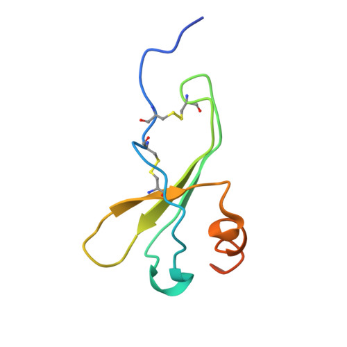
Crystal Structures of Oligomeric Forms of the Ip-10/Cxcl10 Chemokine

Swaminathan, G.J.Holloway, D.E.Colvin, R.A.Campanella, G.K.Papageorgiou, A.C.Luster, A.D.Acharya, K.R.

(2003) Structure 11: 521

  • DOI: https://doi.org/10.1016/s0969-2126(03)00070-4
  • Primary Citation of Related Structures:  
    1O7Y, 1O7Z, 1O80

  • PubMed Abstract: 

    We have determined the structure of wild-type IP-10 from three crystal forms. The crystals provide eight separate models of the IP-10 chain, all differing substantially from a monomeric IP-10 variant examined previously by NMR spectroscopy. In each crystal form, IP-10 chains form conventional beta sheet dimers, which, in turn, form a distinct tetrameric assembly. The M form tetramer is reminiscent of platelet factor 4, whereas the T and H forms feature a novel twelve-stranded beta sheet. Analytical ultracentrifugation indicates that, in free solution, IP-10 exists in a monomer-dimer equilibrium with a dissociation constant of 9 microM. We propose that the tetrameric structures may represent species promoted by the binding of glycosaminoglycans. The binding sites for several IP-10-neutralizing mAbs have also been mapped.


  • Organizational Affiliation

    Department of Biology and Biochemistry, University of Bath, Claverton Down, Bath BA2 7AY, United Kingdom.


Macromolecules
Find similar proteins by:  (by identity cutoff)  |  3D Structure
Entity ID: 1
MoleculeChains Sequence LengthOrganismDetailsImage
SMALL INDUCIBLE CYTOKINE B10
A, B, C, D
77Homo sapiensMutation(s): 0 
UniProt & NIH Common Fund Data Resources
Find proteins for P02778 (Homo sapiens)
Explore P02778 
Go to UniProtKB:  P02778
PHAROS:  P02778
GTEx:  ENSG00000169245 
Entity Groups  
Sequence Clusters30% Identity50% Identity70% Identity90% Identity95% Identity100% Identity
UniProt GroupP02778
Sequence Annotations
Expand
  • Reference Sequence
Small Molecules
Ligands 1 Unique
IDChains Name / Formula / InChI Key2D Diagram3D Interactions
SO4
Query on SO4

Download Ideal Coordinates CCD File 
E [auth A],
F [auth C]
SULFATE ION
O4 S
QAOWNCQODCNURD-UHFFFAOYSA-L
Experimental Data & Validation

Experimental Data

  • Method: X-RAY DIFFRACTION
  • Resolution: 3.00 Å
  • R-Value Free: 0.309 
  • R-Value Work: 0.267 
  • R-Value Observed: 0.267 
  • Space Group: C 1 2 1
Unit Cell:
Length ( Å )Angle ( ˚ )
a = 138.979α = 90
b = 53.722β = 105.72
c = 53.366γ = 90
Software Package:
Software NamePurpose
CNSrefinement
DENZOdata reduction
SCALEPACKdata scaling
AMoREphasing

Structure Validation

View Full Validation Report


Entry History 

Deposition Data

Revision History  (Full details and data files)

  • Version 1.0: 2003-05-08
    Type: Initial release
  • Version 1.1: 2011-05-08
    Changes: Version format compliance
  • Version 1.2: 2011-07-13
    Changes: Version format compliance
  • Version 1.3: 2019-07-24
    Changes: Data collection
  • Version 1.4: 2023-12-13
    Changes: Data collection, Database references, Refinement description