1O7I

Crystal structure of a single stranded DNA binding protein


Experimental Data Snapshot

  • Method: X-RAY DIFFRACTION
  • Resolution: 1.20 Å
  • R-Value Free: 0.219 
  • R-Value Work: 0.191 
  • R-Value Observed: 0.193 

wwPDB Validation   3D Report Full Report


This is version 1.4 of the entry. See complete history


Literature

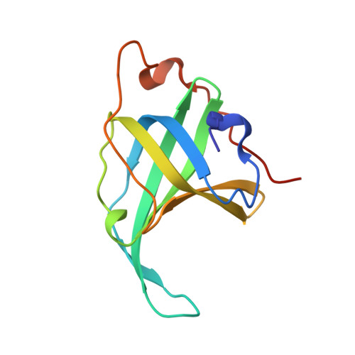
Insights into ssDNA recognition by the OB fold from a structural and thermodynamic study of Sulfolobus SSB protein.

Kerr, I.D.Wadsworth, R.I.Cubeddu, L.Blankenfeldt, W.Naismith, J.H.White, M.F.

(2003) EMBO J 22: 2561-2570

  • DOI: https://doi.org/10.1093/emboj/cdg272
  • Primary Citation of Related Structures:  
    1O7I

  • PubMed Abstract: 

    Information processing pathways such as DNA replication are conserved in eukaryotes and archaea and are significantly different from those found in bacteria. Single-stranded DNA-binding (SSB) proteins (or replication protein A, RPA, in eukaryotes) play a central role in many of these pathways. However, whilst euryarchaea have a eukaryotic-type RPA homologue, crenarchaeal SSB proteins appear much more similar to the bacterial proteins, with a single OB fold for DNA binding and a flexible C-terminal tail that is implicated in protein-protein interactions. We have determined the crystal structure of the SSB protein from the crenarchaeote Sulfolobus solfataricus to 1.26 A. The structure shows a striking and unexpected similarity to the DNA-binding domains of human RPA, providing confirmation of the close relationship between archaea and eukaryotes. The high resolution of the structure, together with thermodynamic and mutational studies of DNA binding, allow us to propose a molecular basis for DNA binding and define the features required for eukaryotic and archaeal OB folds.


  • Organizational Affiliation

    Centre for Biomolecular Science, St Andrews University, Fife, KY16 9ST, UK.


Macromolecules
Find similar proteins by:  (by identity cutoff)  |  3D Structure
Entity ID: 1
MoleculeChains Sequence LengthOrganismDetailsImage
SINGLE STRANDED DNA BINDING PROTEIN
A, B
119Saccharolobus solfataricus P2Mutation(s): 0 
UniProt
Find proteins for Q97W73 (Saccharolobus solfataricus (strain ATCC 35092 / DSM 1617 / JCM 11322 / P2))
Explore Q97W73 
Go to UniProtKB:  Q97W73
Entity Groups  
Sequence Clusters30% Identity50% Identity70% Identity90% Identity95% Identity100% Identity
UniProt GroupQ97W73
Sequence Annotations
Expand
  • Reference Sequence
Experimental Data & Validation

Experimental Data

  • Method: X-RAY DIFFRACTION
  • Resolution: 1.20 Å
  • R-Value Free: 0.219 
  • R-Value Work: 0.191 
  • R-Value Observed: 0.193 
  • Space Group: P 61
Unit Cell:
Length ( Å )Angle ( ˚ )
a = 75.805α = 90
b = 75.805β = 90
c = 70.118γ = 120
Software Package:
Software NamePurpose
REFMACrefinement
MOSFLMdata reduction
SCALAdata scaling
SOLVEphasing

Structure Validation

View Full Validation Report


Entry History 

Deposition Data

Revision History  (Full details and data files)

  • Version 1.0: 2003-06-25
    Type: Initial release
  • Version 1.1: 2011-05-08
    Changes: Version format compliance
  • Version 1.2: 2011-07-13
    Changes: Version format compliance
  • Version 1.3: 2019-01-30
    Changes: Data collection, Experimental preparation, Other
  • Version 1.4: 2019-02-27
    Changes: Data collection, Database references, Experimental preparation