1O70

Novel Fold Revealed by the Structure of a FAS1 Domain Pair from the Insect Cell Adhesion Molecule Fasciclin I


Experimental Data Snapshot

  • Method: X-RAY DIFFRACTION
  • Resolution: 2.60 Å
  • R-Value Free: 0.259 
  • R-Value Work: 0.225 
  • R-Value Observed: 0.225 

wwPDB Validation   3D Report Full Report


This is version 2.0 of the entry. See complete history


Literature

Novel Fold Revealed by the Structure of a Fas1 Domain Pair from the Insect Cell Adhesion Molecule Fasciclin I

Clout, N.J.Tisi, D.Hohenester, E.

(2003) Structure 11: 197

  • DOI: https://doi.org/10.1016/s0969-2126(03)00002-9
  • Primary Citation of Related Structures:  
    1O70

  • PubMed Abstract: 

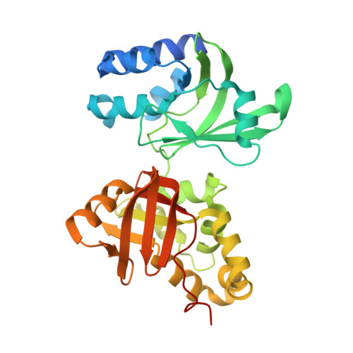
    Fasciclin I is an insect neural cell adhesion molecule consisting of four FAS1 domains, homologs of which are present in many bacterial, plant, and animal proteins. The crystal structure of FAS1 domains 3 and 4 of Drosophila fasciclin I reveals a novel domain fold, consisting of a seven-stranded beta wedge and a number of alpha helices. The two domains are arranged in a linear fashion and interact through a substantial polar interface. Missense mutations in the FAS1 domains of the human protein betaig-h3 cause corneal dystrophies. Many mutations alter highly conserved core residues, but the two most common mutations, affecting Arg-124 and Arg-555, map to exposed alpha-helical regions, suggesting reduced protein solubility as the disease mechanism.


  • Organizational Affiliation

    Biophysics Section, Department of Biological Sciences, Imperial College, SW7 2AZ, London, United Kingdom.


Macromolecules
Find similar proteins by:  (by identity cutoff)  |  3D Structure
Entity ID: 1
MoleculeChains Sequence LengthOrganismDetailsImage
FASCICLIN I324Drosophila melanogasterMutation(s): 0 
UniProt
Find proteins for P10674 (Drosophila melanogaster)
Explore P10674 
Go to UniProtKB:  P10674
Entity Groups  
Sequence Clusters30% Identity50% Identity70% Identity90% Identity95% Identity100% Identity
UniProt GroupP10674
Sequence Annotations
Expand
  • Reference Sequence
Oligosaccharides

Help

Entity ID: 2
MoleculeChains Length2D Diagram Glycosylation3D Interactions
2-acetamido-2-deoxy-beta-D-glucopyranose-(1-4)-2-acetamido-2-deoxy-beta-D-glucopyranose
B
2N-Glycosylation
Glycosylation Resources
GlyTouCan:  G42666HT
GlyCosmos:  G42666HT
GlyGen:  G42666HT
Experimental Data & Validation

Experimental Data

  • Method: X-RAY DIFFRACTION
  • Resolution: 2.60 Å
  • R-Value Free: 0.259 
  • R-Value Work: 0.225 
  • R-Value Observed: 0.225 
  • Space Group: P 43 3 2
Unit Cell:
Length ( Å )Angle ( ˚ )
a = 150.806α = 90
b = 150.806β = 90
c = 150.806γ = 90
Software Package:
Software NamePurpose
CNSrefinement
MOSFLMdata reduction
SCALAdata scaling
MLPHAREphasing

Structure Validation

View Full Validation Report


Entry History 

Deposition Data

Revision History  (Full details and data files)

  • Version 1.0: 2003-02-13
    Type: Initial release
  • Version 1.1: 2012-01-18
    Changes: Derived calculations, Non-polymer description, Other, Refinement description, Version format compliance
  • Version 2.0: 2020-07-29
    Type: Remediation
    Reason: Carbohydrate remediation
    Changes: Atomic model, Data collection, Derived calculations, Other, Structure summary