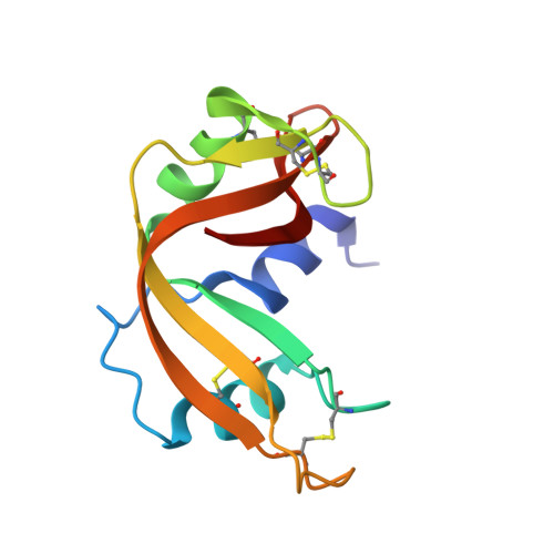
High-resolution crystal structures of ribonuclease A complexed with adenylic and uridylic nucleotide inhibitors. Implications for structure-based design of ribonucleolytic inhibitors
Leonidas, D.D., Chavali, G.B., Oikonomakos, N.G., Chrysina, E.D., Kosmopoulou, M.N., Vlassi, M., Frankling, C., Acharya, K.R.(2003) Protein Sci 12: 2559-2574
- PubMed: 14573867
- DOI: https://doi.org/10.1110/ps.03196603
- Primary Citation of Related Structures:
1O0F, 1O0H, 1O0M, 1O0N, 1O0O - PubMed Abstract:
The crystal structures of bovine pancreatic ribonuclease A (RNase A) in complex with 3',5'-ADP, 2',5'-ADP, 5'-ADP, U-2'-p and U-3'-p have been determined at high resolution. The structures reveal that each inhibitor binds differently in the RNase A active site by anchoring a phosphate group in subsite P1. The most potent inhibitor of all five, 5'-ADP (Ki = 1.2 microM), adopts a syn conformation (in contrast to 3',5'-ADP and 2',5'-ADP, which adopt an anti), and it is the beta- rather than the alpha-phosphate group that binds to P1. 3',5'-ADP binds with the 5'-phosphate group in P1 and the adenosine in the B2 pocket. Two different binding modes are observed in the two RNase A molecules of the asymmetric unit for 2',5'-ADP. This inhibitor binds with either the 3' or the 5' phosphate groups in subsite P1, and in each case, the adenosine binds in two different positions within the B2 subsite. The two uridilyl inhibitors bind similarly with the uridine moiety in the B1 subsite but the placement of a different phosphate group in P1 (2' versus 3') has significant implications on their potency against RNase A. Comparative structural analysis of the RNase A, eosinophil-derived neurotoxin (EDN), eosinophil cationic protein (ECP), and human angiogenin (Ang) complexes with these and other phosphonucleotide inhibitors provides a wealth of information for structure-based design of inhibitors specific for each RNase. These inhibitors could be developed to therapeutic agents that could control the biological activities of EDN, ECP, and ANG, which play key roles in human pathologies.
- Institute of Organic and Pharmaceutical Chemistry, The National Hellenic Research Foundation, 11635 Athens, Greece. ddl@eie.gr
Organizational Affiliation:


