1NYU

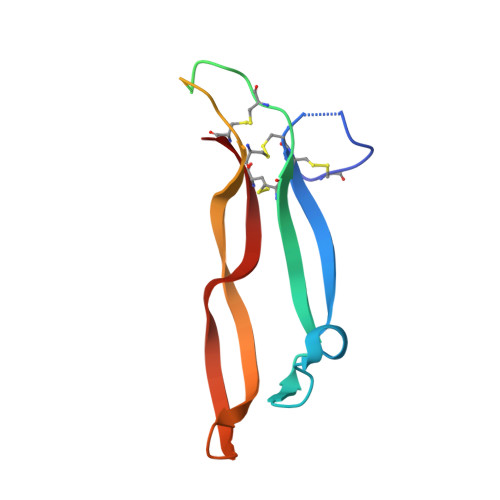
Crystal Structure of Activin A Bound to the ECD of ActRIIB


Experimental Data Snapshot

  • Method: X-RAY DIFFRACTION
  • Resolution: 3.10 Å
  • R-Value Free: 0.291 
  • R-Value Work: 0.268 
  • R-Value Observed: 0.268 

Starting Model: experimental
View more details

wwPDB Validation   3D Report Full Report


This is version 1.5 of the entry. See complete history


Literature

Structures of an ActRIIB:activin A complex reveal a novel binding mode for TGF-beta ligand:receptor interactions

Thompson, T.B.Woodruff, T.K.Jardetzky, T.S.

(2003) EMBO J 22: 1555-1566

  • DOI: https://doi.org/10.1093/emboj/cdg156
  • Primary Citation of Related Structures:  
    1NYS, 1NYU

  • PubMed Abstract: 

    The TGF-beta superfamily of ligands and receptors stimulate cellular events in diverse processes ranging from cell fate specification in development to immune suppression. Activins define a major subgroup of TGF-beta ligands that regulate cellular differentiation, proliferation, activation and apoptosis. Activins signal through complexes formed with type I and type II serine/threonine kinase receptors. We have solved the crystal structure of activin A bound to the extracellular domain of a type II receptor, ActRIIB, revealing the details of this interaction. ActRIIB binds to the outer edges of the activin finger regions, with the two receptors juxtaposed in close proximity, in a mode that differs from TGF-beta3 binding to type II receptors. The dimeric activin A structure differs from other known TGF-beta ligand structures, adopting a compact folded-back conformation. The crystal structure of the complex is consistent with recruitment of two type I receptors into a close packed arrangement at the cell surface and suggests that diversity in the conformational arrangements of TGF-beta ligand dimers could influence cellular signaling processes.


  • Organizational Affiliation

    Department of Biochemistry, Northwestern University, 2205 Tech Drive, Evanston, IL 60208, USA.


Macromolecules
Find similar proteins by:  (by identity cutoff)  |  3D Structure
Entity ID: 1
MoleculeChains Sequence LengthOrganismDetailsImage
activin receptor
A, C
105Rattus norvegicusMutation(s): 0 
Gene Names: actrIIb
UniProt
Find proteins for P38445 (Rattus norvegicus)
Explore P38445 
Go to UniProtKB:  P38445
Entity Groups  
Sequence Clusters30% Identity50% Identity70% Identity90% Identity95% Identity100% Identity
UniProt GroupP38445
Sequence Annotations
Expand
  • Reference Sequence
Find similar proteins by:  (by identity cutoff)  |  3D Structure
Entity ID: 2
MoleculeChains Sequence LengthOrganismDetailsImage
Inhibin beta A chain
B, D
116Homo sapiensMutation(s): 0 
Gene Names: INHBA
UniProt & NIH Common Fund Data Resources
Find proteins for P08476 (Homo sapiens)
Explore P08476 
Go to UniProtKB:  P08476
PHAROS:  P08476
GTEx:  ENSG00000122641 
Entity Groups  
Sequence Clusters30% Identity50% Identity70% Identity90% Identity95% Identity100% Identity
UniProt GroupP08476
Sequence Annotations
Expand
  • Reference Sequence
Experimental Data & Validation

Experimental Data

  • Method: X-RAY DIFFRACTION
  • Resolution: 3.10 Å
  • R-Value Free: 0.291 
  • R-Value Work: 0.268 
  • R-Value Observed: 0.268 
  • Space Group: P 41 21 2
Unit Cell:
Length ( Å )Angle ( ˚ )
a = 141.33α = 90
b = 141.33β = 90
c = 46.021γ = 90
Software Package:
Software NamePurpose
CNSrefinement
MAR345data collection
SCALEPACKdata scaling
BRUTEphasing
BEASTphasing
CNSphasing

Structure Validation

View Full Validation Report


Entry History 

Deposition Data

Revision History  (Full details and data files)

  • Version 1.0: 2003-04-08
    Type: Initial release
  • Version 1.1: 2008-04-29
    Changes: Version format compliance
  • Version 1.2: 2011-07-13
    Changes: Version format compliance
  • Version 1.3: 2012-05-30
    Changes: Database references
  • Version 1.4: 2017-10-11
    Changes: Refinement description
  • Version 1.5: 2023-08-16
    Changes: Data collection, Database references, Refinement description