1NUC | pdb_00001nuc

STAPHYLOCOCCAL NUCLEASE, V23C VARIANT


Experimental Data Snapshot

  • Method: X-RAY DIFFRACTION
  • Resolution: 1.90 Å
  • R-Value Work: 
    0.173 (Depositor) 
  • R-Value Observed: 
    0.173 (Depositor) 

Starting Model: experimental
View more details

wwPDB Validation 3D Report Full Report

Validation slider image for 1NUC

This is version 1.5 of the entry. See complete history

Literature

Mobile unnatural amino acid side chains in the core of staphylococcal nuclease.

Wynn, R.Harkins, P.C.Richards, F.M.Fox, R.O.

(1996) Protein Sci 5: 1026-1031

  • DOI: https://doi.org/10.1002/pro.5560050605
  • Primary Citation Related Structures: 
    1A2T, 1A2U, 1AEX, 1NUC, 2NUC, 3NUC, 5NUC

  • PubMed Abstract: 

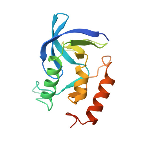
    The structures of several variants of staphylococcal nuclease with long flexible unnatural amino acid side chains in the hydrophobic core have been determined by X-ray crystallography. The unnatural amino acids are disulfide moieties between the lone cysteine residue in V23C nuclease and methane, ethane, 1-n-propane, 1-n-butane, 1-n-pentane, and 2-hydroxyethyl thiols. We have examined changes in the core packing of these mutants. Side chains as large as the 1-n-propyl cysteine disulfide can be incorporated without perturbation of the structure. This is due, in part, to cavities present in the wild-type protein. The longest side chains are not well defined, even though they remain buried within the protein interior. These results suggest that the enthalpy-entropy balance that governs the rigidity of protein interiors favors tight packing only weakly. Additionally, the tight packing observed normally in protein interiors may reflect, in part, the limited numbers of rotamers available to the natural amino acids.


  • Organizational Affiliation
    • Department of Molecular Biophysics and Biochemistry, Yale University, New Haven, Connecticut 06511, USA. wynnr@al.lldmpc.umc.dupont.com

Macromolecule Content 

  • Total Structure Weight: 17.29 kDa 
  • Atom Count: 1,152 
  • Modeled Residue Count: 135 
  • Deposited Residue Count: 149 
  • Unique protein chains: 1

Macromolecules

Find similar proteins by:|  3D Structure
Entity ID: 1
MoleculeChains  Sequence LengthOrganismDetailsImage
STAPHYLOCOCCAL NUCLEASE149Staphylococcus aureusMutation(s): 1 
EC: 3.1.31.1
UniProt
Find proteins for P00644 (Staphylococcus aureus)
Explore P00644 
Go to UniProtKB:  P00644
Entity Groups
Sequence Clusters30% Identity50% Identity70% Identity90% Identity95% Identity100% Identity
UniProt GroupP00644
Sequence Annotations
Expand
Reference Sequence

Small Molecules

Ligands 2 Unique
IDChains Name / Formula / InChI Key2D Diagram3D Interactions
THP

Query on THP


Download:Ideal Coordinates CCD File
C [auth A]THYMIDINE-3',5'-DIPHOSPHATE
C10 H16 N2 O11 P2
CSNCBOPUCJOHLS-XLPZGREQSA-N
CA

Query on CA


Download:Ideal Coordinates CCD File
B [auth A]CALCIUM ION
Ca
BHPQYMZQTOCNFJ-UHFFFAOYSA-N

Experimental Data & Validation

Experimental Data

  • Method: X-RAY DIFFRACTION
  • Resolution: 1.90 Å
  • R-Value Work:  0.173 (Depositor) 
  • R-Value Observed: 0.173 (Depositor) 
Space Group: P 41
Unit Cell:
Length ( Å )Angle ( ˚ )
a = 47.4α = 90
b = 47.4β = 90
c = 62.8γ = 90
Software Package:
Software NamePurpose
X-PLORmodel building
X-PLORrefinement
DENZOdata reduction
SCALEPACKdata scaling
X-PLORphasing

Structure Validation

View Full Validation Report


Entry History 

Deposition Data

Revision History  (Full details and data files)

  • Version 1.0: 1997-06-16
    Type: Initial release
  • Version 1.1: 2008-03-24
    Changes: Version format compliance
  • Version 1.2: 2011-07-13
    Changes: Version format compliance
  • Version 1.3: 2021-11-03
    Changes: Database references, Derived calculations, Other
  • Version 1.4: 2023-08-09
    Changes: Refinement description
  • Version 1.5: 2024-05-22
    Changes: Data collection