1NRV | pdb_00001nrv

Crystal structure of the SH2 domain of Grb10


Experimental Data Snapshot

  • Method: X-RAY DIFFRACTION
  • Resolution: 1.65 Å
  • R-Value Free: 
    0.242 (Depositor), 0.243 (DCC) 
  • R-Value Work: 
    0.224 (Depositor), 0.222 (DCC) 
  • R-Value Observed: 
    0.224 (Depositor) 

Starting Model: experimental
View more details

wwPDB Validation 3D Report Full Report

Validation slider image for 1NRV

This is version 1.3 of the entry. See complete history

Literature

Structural basis for dimerization of the Grb10 Src homology 2 domain. Implications for ligand specificity.

Stein, E.G.Ghirlando, R.Hubbard, S.R.

(2003) J Biological Chem 278: 13257-13264

  • DOI: https://doi.org/10.1074/jbc.M212026200
  • Primary Citation Related Structures: 
    1NRV

  • PubMed Abstract: 

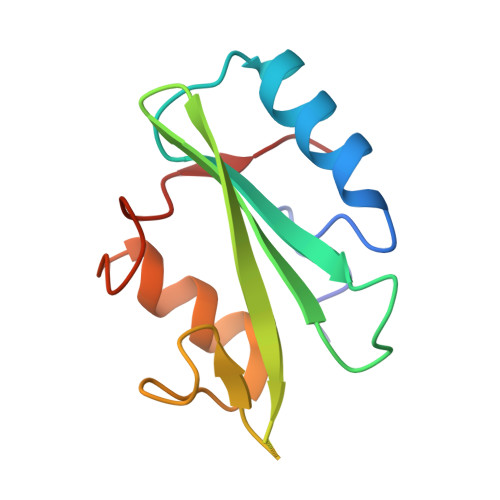
    Grb7, Grb10, and Grb14 are members of a distinct family of adapter proteins that interact with various receptor tyrosine kinases upon receptor activation. Proteins in this family contain several modular signaling domains including a pleckstrin homology (PH) domain, a BPS (between PH and SH2) domain, and a C-terminal Src homology 2 (SH2) domain. Although SH2 domains are typically monomeric, we show that the Grb10 SH2 domain and also full-length Grb10 gamma are dimeric in solution under physiologic conditions. The crystal structure of the Grb10 SH2 domain at 1.65-A resolution reveals a non-covalent dimer whose interface comprises residues within and flanking the C-terminal alpha helix, which are conserved in the Grb7/Grb10/Grb14 family but not in other SH2 domains. Val-522 in the BG loop (BG3) and Asp-500 in the EF loop (EF1) are positioned to interfere with the binding of the P+3 residue of a phosphopeptide ligand. These structural features of the Grb10 SH2 domain will favor binding of dimeric, turn-containing phosphotyrosine sequences, such as the phosphorylated activation loops in the two beta subunits of the insulin and insulin-like growth factor-1 receptors. Moreover, the structure suggests the mechanism by which the Grb7 SH2 domain binds selectively to pTyr-1139 (pYVNQ) in Her2, which along with Grb7 is co-amplified in human breast cancers.


  • Organizational Affiliation
    • Skirball Institute of Biomolecular Medicine and Department of Pharmacology, New York University School of Medicine, New York, New York 10016, USA.

Macromolecule Content 

  • Total Structure Weight: 24.72 kDa 
  • Atom Count: 1,843 
  • Modeled Residue Count: 200 
  • Deposited Residue Count: 210 
  • Unique protein chains: 1

Macromolecules

Find similar proteins by:|  3D Structure
Entity ID: 1
MoleculeChains  Sequence LengthOrganismDetailsImage
Growth factor receptor-bound protein 10
A, B
105Homo sapiensMutation(s): 0 
Gene Names: GRB10 OR GRBIR OR KIAA0207
UniProt & NIH Common Fund Data Resources
Find proteins for Q13322 (Homo sapiens)
Explore Q13322 
Go to UniProtKB:  Q13322
PHAROS:  Q13322
GTEx:  ENSG00000106070 
Entity Groups
Sequence Clusters30% Identity50% Identity70% Identity90% Identity95% Identity100% Identity
UniProt GroupQ13322
Sequence Annotations
Expand
Reference Sequence

Experimental Data & Validation

Experimental Data

  • Method: X-RAY DIFFRACTION
  • Resolution: 1.65 Å
  • R-Value Free:  0.242 (Depositor), 0.243 (DCC) 
  • R-Value Work:  0.224 (Depositor), 0.222 (DCC) 
  • R-Value Observed: 0.224 (Depositor) 
Space Group: P 1 21 1
Unit Cell:
Length ( Å )Angle ( ˚ )
a = 28.82α = 90
b = 49.04β = 96.62
c = 79.61γ = 90
Software Package:
Software NamePurpose
DENZOdata reduction
SCALEPACKdata scaling
AMoREphasing
CNSrefinement

Structure Validation

View Full Validation Report


Entry History 

Deposition Data

Revision History  (Full details and data files)

  • Version 1.0: 2003-04-15
    Type: Initial release
  • Version 1.1: 2008-04-29
    Changes: Version format compliance
  • Version 1.2: 2011-07-13
    Changes: Version format compliance
  • Version 1.3: 2023-08-16
    Changes: Data collection, Database references, Refinement description