1NQL

Structure of the extracellular domain of human epidermal growth factor (EGF) receptor in an inactive (low pH) complex with EGF.


Experimental Data Snapshot

  • Method: X-RAY DIFFRACTION
  • Resolution: 2.80 Å
  • R-Value Free: 0.310 
  • R-Value Work: 0.241 
  • R-Value Observed: 0.247 

wwPDB Validation   3D Report Full Report


This is version 2.1 of the entry. See complete history


Literature

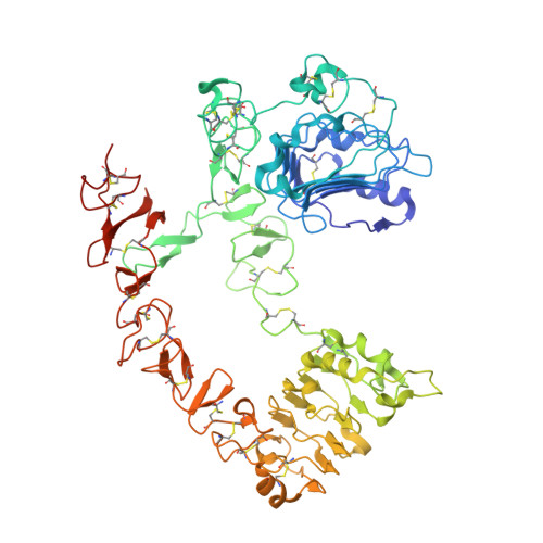
EGF activates its receptor by removing interactions that auto-inhibit ectodomain dimerization

Ferguson, K.M.Berger, M.B.Mendrola, J.M.Cho, H.Leahy, D.J.Lemmon, M.A.

(2003) Mol Cell 11: 507-517

  • DOI: https://doi.org/10.1016/s1097-2765(03)00047-9
  • Primary Citation of Related Structures:  
    1NQL

  • PubMed Abstract: 
  • Epidermal growth factor (EGF) receptor is the prototype of the ErbB (HER) family receptor tyrosine kinases (RTKs), which regulate cell growth and differentiation and are implicated in many human cancers. EGF activates its receptor by inducing dimerization of the 621 amino acid EGF receptor extracellular region ...

    Epidermal growth factor (EGF) receptor is the prototype of the ErbB (HER) family receptor tyrosine kinases (RTKs), which regulate cell growth and differentiation and are implicated in many human cancers. EGF activates its receptor by inducing dimerization of the 621 amino acid EGF receptor extracellular region. We describe the 2.8 A resolution crystal structure of this entire extracellular region (sEGFR) in an unactivated state. The structure reveals an autoinhibited configuration, where the dimerization interface recently identified in activated sEGFR structures is completely occluded by intramolecular interactions. To activate the receptor, EGF binding must promote a large domain rearrangement that exposes this dimerization interface. This contrasts starkly with other RTK activation mechanisms and suggests new approaches for designing ErbB receptor antagonists.


    Organizational Affiliation

    Department of Biochemistry and Biophysics, University of Pennsylvania School of Medicine, Philadelphia, PA 19104, USA. fertuso2@mail.med.upenn.edu


Macromolecules
Find similar proteins by:  (by identity cutoff)  |  3D Structure
Entity ID: 1
MoleculeChainsSequence LengthOrganismDetailsImage
epidermal growth factor receptor624Homo sapiensMutation(s): 0 
Gene Names: EGFRERBBERBB1HER1
EC: 2.7.10.1
UniProt & NIH Common Fund Data Resources
Find proteins for P00533 (Homo sapiens)
Explore P00533 
Go to UniProtKB:  P00533
PHAROS:  P00533
Entity Groups  
Sequence Clusters30% Identity50% Identity70% Identity90% Identity95% Identity100% Identity
UniProt GroupP00533
Protein Feature View
Expand
  • Reference Sequence
Find similar proteins by:  (by identity cutoff)  |  3D Structure
Entity ID: 2
MoleculeChainsSequence LengthOrganismDetailsImage
epidermal growth factor53Homo sapiensMutation(s): 0 
Gene Names: EGF
UniProt & NIH Common Fund Data Resources
Find proteins for P01133 (Homo sapiens)
Explore P01133 
Go to UniProtKB:  P01133
PHAROS:  P01133
Entity Groups  
Sequence Clusters30% Identity50% Identity70% Identity90% Identity95% Identity100% Identity
UniProt GroupP01133
Protein Feature View
Expand
  • Reference Sequence
Oligosaccharides

Help

Entity ID: 3
MoleculeChainsChain Length2D DiagramGlycosylation3D Interactions
beta-D-mannopyranose-(1-4)-2-acetamido-2-deoxy-beta-D-glucopyranose-(1-4)-2-acetamido-2-deoxy-beta-D-glucopyranose
C, D
3N-Glycosylation Oligosaccharides Interaction
Glycosylation Resources
GlyTouCan:  G15407YE
GlyCosmos:  G15407YE
GlyGen:  G15407YE
Entity ID: 4
MoleculeChainsChain Length2D DiagramGlycosylation3D Interactions
2-acetamido-2-deoxy-beta-D-glucopyranose-(1-4)-2-acetamido-2-deoxy-beta-D-glucopyranose
E
2N-Glycosylation Oligosaccharides Interaction
Glycosylation Resources
GlyTouCan:  G42666HT
GlyCosmos:  G42666HT
GlyGen:  G42666HT
Small Molecules
Ligands 1 Unique
IDChainsName / Formula / InChI Key2D Diagram3D Interactions
NAG
Query on NAG

Download Ideal Coordinates CCD File 
F [auth A],
G [auth A],
H [auth A],
I [auth A],
J [auth A]
2-acetamido-2-deoxy-beta-D-glucopyranose
C8 H15 N O6
OVRNDRQMDRJTHS-FMDGEEDCSA-N
 Ligand Interaction
Experimental Data & Validation

Experimental Data

  • Method: X-RAY DIFFRACTION
  • Resolution: 2.80 Å
  • R-Value Free: 0.310 
  • R-Value Work: 0.241 
  • R-Value Observed: 0.247 
  • Space Group: C 1 2 1
Unit Cell:
Length ( Å )Angle ( ˚ )
a = 119.17α = 90
b = 103.66β = 119.27
c = 101.49γ = 90
Software Package:
Software NamePurpose
REFMACrefinement
MOSFLMdata reduction
CCP4data scaling
AMoREphasing

Structure Validation

View Full Validation Report


Entry History 

Deposition Data

Revision History  (Full details and data files)

  • Version 1.0: 2003-03-11
    Type: Initial release
  • Version 1.1: 2008-04-29
    Changes: Version format compliance
  • Version 1.2: 2011-07-13
    Changes: Non-polymer description, Version format compliance
  • Version 2.0: 2020-07-29
    Type: Remediation
    Reason: Carbohydrate remediation
    Changes: Advisory, Atomic model, Data collection, Derived calculations, Structure summary
  • Version 2.1: 2023-08-16
    Changes: Advisory, Data collection, Database references, Refinement description, Structure summary