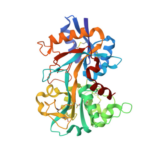
Structural evidence for a pH-sensitive dilysine trigger in the hen ovotransferrin N-lobe: implications for transferrin iron release.
Dewan, J.C., Mikami, B., Hirose, M., Sacchettini, J.C.(1993) Biochemistry 32: 11963-11968
- PubMed: 8218271
- DOI: https://doi.org/10.1021/bi00096a004
- Primary Citation of Related Structures:
1NNT - PubMed Abstract:
Members of the transferrin family of proteins are involved in Fe3+ transport (serum transferrins) and are also believed to possess antimicrobial activity (ovotransferrins and lactoferrins). The structure of the monoferric N-terminal half-molecule of hen ovotransferrin, reported here at 2.3-A resolution, reveals an unusual interdomain interaction formed between the side-chain NZ atoms of Lys 209 and Lys 301, which are 2.3 A apart. This strong interaction appears to be an example of a low-barrier hydrogen bond between the two lysine NZ atoms, both of which are also involved in a hydrogen-bonding interaction with the aromatic ring of a tyrosine residue. Crystals of the protein were grown at pH 5.9, which is well below the usual pKa approximately 10 for a lysine side chain. We suggest that the pKa of either one or both of these residues lies below the pH of the structure determination and is, therefore, not positively charged. This finding may serve to explain, on a molecular basis, the pH dependence of transferrin Fe3+ release. We propose that uptake of the Fe(3+)-transferrin complex into an acidic endosome (viz., pH approximately 5.0) via receptor-mediated endocytosis will result in the protonation of both lysine residues. The close proximity of the two resulting positive charges, and their location on opposite domains of the N-lobe, might well be the driving force that opens the two domains of the protein, exposing the Fe3+ ion and facilitating its release.(ABSTRACT TRUNCATED AT 250 WORDS)
- Department of Biochemistry, Albert Einstein College of Medicine, Bronx, New York 10461.
Organizational Affiliation:


