Structure in solution of the major cold-shock protein from Bacillus subtilis.
Schnuchel, A., Wiltscheck, R., Czisch, M., Herrler, M., Willimsky, G., Graumann, P., Marahiel, M.A., Holak, T.A.(1993) Nature 364: 169-171
- PubMed: 8321289 Search on PubMed
- DOI: https://doi.org/10.1038/364169a0
- Primary Citation Related Structures:
1NMF, 1NMG - PubMed Abstract:
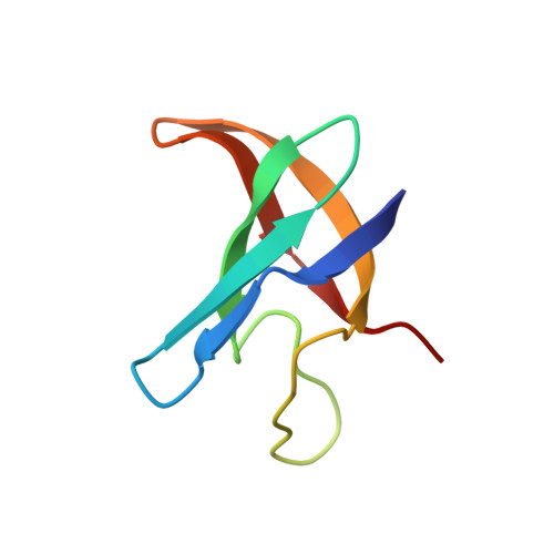
The cold-shock domain (CSD) is found in many eukaryotic transcriptional factors and is responsible for the specific binding to DNA of a cis-element called the Y-box. The same domain exists in the sequence of the Xenopus RNA-binding proteins FRG Y1 and FRG Y2 (refs 1, 3). The major cold-shock proteins of Escherichia coli (CS7.4) and B. subtilis (CspB) have sequences that are more than 40 per cent identical to the cold-shock domain. We present here the three-dimensional structure of CspB determined by nuclear magnetic resonance spectroscopy. The 67-residue protein consists of an antiparallel five-stranded beta-barrel with strands connected by turns and loops. The structure resembles that of staphylococcal nuclease and the gene-5 single-stranded-DNA-binding protein. A three-stranded beta-sheet, which contains the conserved RNA-binding motif RNP1 as well as a motif similar to RNP2 in two neighbouring antiparallel beta-strands, has basic and aromatic residues at its surface which could serve as a binding site for single-stranded DNA. CspB binds to single-stranded DNA in gel retardation experiments.
- Max-Planck-Institut für Biochemie, Martinsried bei München, Germany.
Organizational Affiliation: