1NK1 | pdb_00001nk1

NK1 FRAGMENT OF HUMAN HEPATOCYTE GROWTH FACTOR/SCATTER FACTOR (HGF/SF) AT 2.5 ANGSTROM RESOLUTION


Experimental Data Snapshot

  • Method: X-RAY DIFFRACTION
  • Resolution: 2.50 Å
  • R-Value Free: 
    0.319 (Depositor), 0.317 (DCC) 
  • R-Value Work: 
    0.245 (Depositor), 0.246 (DCC) 
  • R-Value Observed: 
    0.245 (Depositor) 

Starting Models: experimental
View more details

wwPDB Validation 3D Report Full Report

Validation slider image for 1NK1

This is version 1.5 of the entry. See complete history

Literature

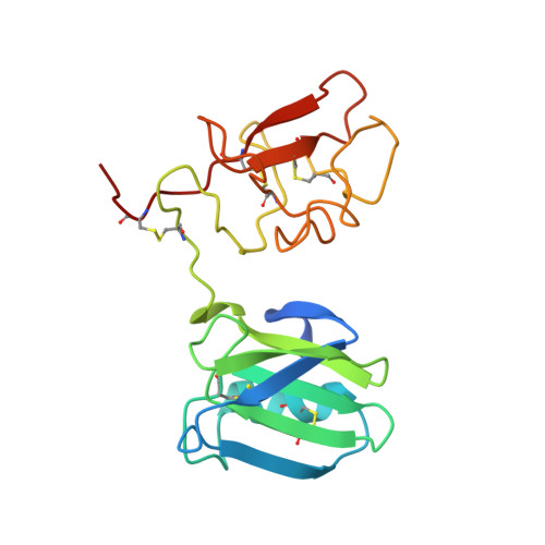
Crystal structure of the NK1 fragment of HGF/SF suggests a novel mode for growth factor dimerization and receptor binding.

Chirgadze, D.Y.Hepple, J.P.Zhou, H.Byrd, R.A.Blundell, T.L.Gherardi, E.

(1999) Nat Struct Biol 6: 72-79

  • DOI: https://doi.org/10.1038/4947
  • Primary Citation Related Structures: 
    1NK1

  • PubMed Abstract: 

    Although ligand-induced receptor dimerization is a common prerequisite for receptor activation, the mode by which different growth factors bind their receptors and cause them to dimerize varies considerably. Here we report the crystal structure at 2.5 A resolution of NK1, a receptor-binding fragment and a natural splice variant of hepatocyte growth factor/scatter factor (HGF/SF). NK1 assembles as a homodimer in the asymmetric unit, revealing a novel mode of growth factor dimerization produced by close packing of the N domain of one subunit and the kringle domain of the other, thus bringing the two linkers in close proximity. The structure suggests the presence of a binding site for heparan sulfate chains and a mechanism by which the NK1 dimer may engage two receptor molecules through clusters of amino acids located on each protomer and on opposite surfaces of the homodimer. We also report that short (14-mer) heparin fragments effectively dimerize NK1 in solution, implying that heparan sulfate chains may stabilize the NK1 dimer. These results provide a basis for the agonistic activity of NK1 and have implications for the mechanism of receptor binding of HGF/SF.


  • Organizational Affiliation
    • Department of Biochemistry, University of Cambridge, UK.

Macromolecule Content 

  • Total Structure Weight: 42.34 kDa 
  • Atom Count: 2,823 
  • Modeled Residue Count: 344 
  • Deposited Residue Count: 366 
  • Unique protein chains: 1

Macromolecules

Find similar proteins by:|  3D Structure
Entity ID: 1
MoleculeChains  Sequence LengthOrganismDetailsImage
PROTEIN (HEPATOCYTE GROWTH FACTOR PRECURSOR)
A, B
183Homo sapiensMutation(s): 1 
Gene Names: HEPATOCYTE GROWTH FACTOR/SCATTER FACTOR
UniProt & NIH Common Fund Data Resources
Find proteins for P14210 (Homo sapiens)
Explore P14210 
Go to UniProtKB:  P14210
PHAROS:  P14210
GTEx:  ENSG00000019991 
Entity Groups
Sequence Clusters30% Identity50% Identity70% Identity90% Identity95% Identity100% Identity
UniProt GroupP14210
Sequence Annotations
Expand
Reference Sequence

Experimental Data & Validation

Experimental Data

  • Method: X-RAY DIFFRACTION
  • Resolution: 2.50 Å
  • R-Value Free:  0.319 (Depositor), 0.317 (DCC) 
  • R-Value Work:  0.245 (Depositor), 0.246 (DCC) 
  • R-Value Observed: 0.245 (Depositor) 
Space Group: P 1 21 1
Unit Cell:
Length ( Å )Angle ( ˚ )
a = 53.42α = 90
b = 63.56β = 96.04
c = 56.81γ = 90
Software Package:
Software NamePurpose
DENZOdata reduction
SCALEPACKdata scaling
AMoREphasing
X-PLORrefinement

Structure Validation

View Full Validation Report


Entry History 

Deposition Data

Revision History  (Full details and data files)

  • Version 1.0: 1999-01-13
    Type: Initial release
  • Version 1.1: 2008-04-27
    Changes: Version format compliance
  • Version 1.2: 2011-07-13
    Changes: Version format compliance
  • Version 1.3: 2021-11-03
    Changes: Database references
  • Version 1.4: 2023-08-16
    Changes: Data collection, Refinement description
  • Version 1.5: 2024-11-06
    Changes: Structure summary