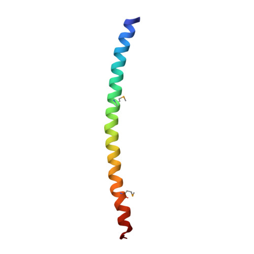
Homotetrameric Structure of the SNAP-23 N-terminal Coiled-coil Domain
Freedman, S.J., Song, H.K., Xu, Y., Sun, Z.Y., Eck, M.J.(2003) J Biol Chem 278: 13462-13467
- PubMed: 12556468 Search on PubMed
- DOI: https://doi.org/10.1074/jbc.M210483200
- Primary Citation Related Structures:
1NHL - PubMed Abstract:
SNARE proteins mediate intracellular membrane fusion by forming a coiled-coil complex to merge opposing membranes. A "fusion-active" neuronal SNARE complex is a parallel four-helix bundle containing two coiled-coil domains from SNAP-25 and one coiled-coil domain each from syntaxin-1a and VAMP-2. "Prefusion" assembly intermediate complexes can also form from these SNAREs. We studied the N-terminal coiled-coil domain of SNAP-23 (SNAP-23N), a non-neuronal homologue of SNAP-25, and its interaction with other coiled-coil domains. SNAP-23N can assemble spontaneously with the coiled-coil domains from SNAP-23C, syntaxin-4, and VAMP-3 to form a heterotetrameric complex. Unexpectedly, pure SNAP-23N crystallizes as a coiled-coil homotetrameric complex. The four helices have a parallel orientation and are symmetrical about the long axis. The complex is stabilized through the interaction of conserved hydrophobic residues comprising the a and d positions of the coiled-coil heptad repeats. In addition, a central, highly conserved glutamine residue (Gln-48) is buried within the interface by hydrogen bonding between glutamine side chains derived from adjacent subunits and to solvent molecules. A comparison of the SNAP-23N structure to other SNARE complex structures reveals how a simple coiled-coil motif can form diverse SNARE complexes.
- Division of Hemostasis and Thrombosis, Beth Israel Deaconess Medical Center, Boston, Massachusetts 02115, USA. sfreedm2@caregroup.harvard.edu
Organizational Affiliation: