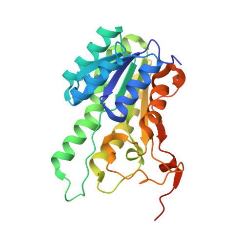
1NFF

Crystal structure of Rv2002 gene product from Mycobacterium tuberculosis


Experimental Data Snapshot

  • Method: X-RAY DIFFRACTION
  • Resolution: 1.80 Å
  • R-Value Free: 0.199 
  • R-Value Work: 0.164 
  • R-Value Observed: 0.164 

wwPDB Validation   3D Report Full Report


Ligand Structure Quality Assessment 


This is version 1.4 of the entry. See complete history


Literature

Directed evolution approach to a structural genomics project: Rv2002 from Mycobacterium tuberculosis

Yang, J.K.Park, M.S.Waldo, G.S.Suh, S.W.

(2003) Proc Natl Acad Sci U S A 100: 455-460

  • DOI: https://doi.org/10.1073/pnas.0137017100
  • Primary Citation of Related Structures:  
    1NFF, 1NFQ, 1NFR

  • PubMed Abstract: 

    One of the serious bottlenecks in structural genomics projects is overexpression of the target proteins in soluble form. We have applied the directed evolution technique and prepared soluble mutants of the Mycobacterium tuberculosis Rv2002 gene product, the wild type of which had been expressed as inclusion bodies in Escherichia coli. A triple mutant I6TV47MT69K (Rv2002-M3) was chosen for structural and functional characterizations. Enzymatic assays indicate that the Rv2002-M3 protein has a high catalytic activity as a NADH-dependent 3alpha, 20beta-hydroxysteroid dehydrogenase. We have determined the crystal structures of a binary complex with NAD(+) and a ternary complex with androsterone and NADH. The structure reveals that Asp-38 determines the cofactor specificity. The catalytic site includes the triad Ser-140Tyr-153Lys-157. Additionally, it has an unusual feature, Glu-142. Enzymatic assays of the E142A mutant of Rv2002-M3 indicate that Glu-142 reverses the effect of Lys-157 in influencing the pKa of Tyr-153. This study suggests that the Rv2002 gene product is a unique member of the SDR family and is likely to be involved in steroid metabolism in M. tuberculosis. Our work demonstrates the power of the directed evolution technique as a general way of overcoming the difficulties in overexpressing the target proteins in soluble form.


  • Organizational Affiliation

    Structural Proteomics Laboratory, School of Chemistry and Molecular Engineering, Seoul National University, Seoul 151-742, South Korea.


Macromolecules
Find similar proteins by:  (by identity cutoff)  |  3D Structure
Entity ID: 1
MoleculeChains Sequence LengthOrganismDetailsImage
Putative oxidoreductase Rv2002
A, B
260Mycobacterium tuberculosisMutation(s): 3 
EC: 1.1.1.53
UniProt
Find proteins for P9WGT1 (Mycobacterium tuberculosis (strain ATCC 25618 / H37Rv))
Explore P9WGT1 
Go to UniProtKB:  P9WGT1
Entity Groups  
Sequence Clusters30% Identity50% Identity70% Identity90% Identity95% Identity100% Identity
UniProt GroupP9WGT1
Sequence Annotations
Expand
  • Reference Sequence
Small Molecules
Ligands 1 Unique
IDChains Name / Formula / InChI Key2D Diagram3D Interactions
NAD
Query on NAD

Download Ideal Coordinates CCD File 
C [auth A],
D [auth B]
NICOTINAMIDE-ADENINE-DINUCLEOTIDE
C21 H27 N7 O14 P2
BAWFJGJZGIEFAR-NNYOXOHSSA-N
Experimental Data & Validation

Experimental Data

  • Method: X-RAY DIFFRACTION
  • Resolution: 1.80 Å
  • R-Value Free: 0.199 
  • R-Value Work: 0.164 
  • R-Value Observed: 0.164 
  • Space Group: P 32 2 1
Unit Cell:
Length ( Å )Angle ( ˚ )
a = 70.377α = 90
b = 70.377β = 90
c = 148.934γ = 120
Software Package:
Software NamePurpose
CNSrefinement
MOSFLMdata reduction
CCP4data scaling
AMoREphasing

Structure Validation

View Full Validation Report


Ligand Structure Quality Assessment 


Entry History 

Deposition Data

Revision History  (Full details and data files)

  • Version 1.0: 2002-12-30
    Type: Initial release
  • Version 1.1: 2008-04-29
    Changes: Version format compliance
  • Version 1.2: 2011-07-13
    Changes: Derived calculations, Version format compliance
  • Version 1.3: 2021-11-10
    Changes: Database references, Derived calculations
  • Version 1.4: 2024-05-29
    Changes: Data collection