1NCG

STRUCTURAL BASIS OF CELL-CELL ADHESION BY CADHERINS


Experimental Data Snapshot

  • Method: X-RAY DIFFRACTION
  • Resolution: 2.10 Å
  • R-Value Free: 0.201 
  • R-Value Work: 0.217 
  • R-Value Observed: 0.217 

wwPDB Validation   3D Report Full Report


This is version 1.4 of the entry. See complete history


Literature

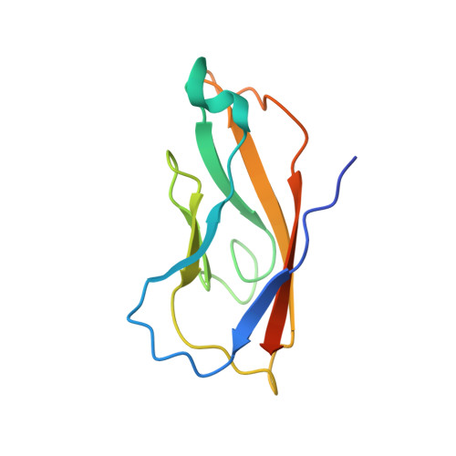
Structural basis of cell-cell adhesion by cadherins.

Shapiro, L.Fannon, A.M.Kwong, P.D.Thompson, A.Lehmann, M.S.Grubel, G.Legrand, J.F.Als-Nielsen, J.Colman, D.R.Hendrickson, W.A.

(1995) Nature 374: 327-337

  • DOI: https://doi.org/10.1038/374327a0
  • Primary Citation of Related Structures:  
    1NCG, 1NCH, 1NCI

  • PubMed Abstract: 

    Crystal structures of the amino-terminal domain of N-cadherin provide a picture at the atomic level of a specific adhesive contact between cells. A repeated set of dimer interfaces is common to the structure in three lattices. These interactions combine to form a linear zipper of molecules that mirrors the linear structure of the intracellular filaments with which cadherins associate. This cell-adhesion zipper may provide a mechanism to marshal individual molecular adhesive interactions into strong bonds between cells.


  • Organizational Affiliation

    Department of Biochemistry and Molecular Biophysics, Columbia University, New York, New York 10032.


Macromolecules
Find similar proteins by:  (by identity cutoff)  |  3D Structure
Entity ID: 1
MoleculeChains Sequence LengthOrganismDetailsImage
N-CADHERIN110Mus musculusMutation(s): 0 
UniProt
Find proteins for P15116 (Mus musculus)
Explore P15116 
Go to UniProtKB:  P15116
Entity Groups  
Sequence Clusters30% Identity50% Identity70% Identity90% Identity95% Identity100% Identity
UniProt GroupP15116
Sequence Annotations
Expand
  • Reference Sequence
Small Molecules
Ligands 1 Unique
IDChains Name / Formula / InChI Key2D Diagram3D Interactions
YB
Query on YB

Download Ideal Coordinates CCD File 
B [auth A]YTTERBIUM (III) ION
Yb
AWSFICBXMUKWSK-UHFFFAOYSA-N
Experimental Data & Validation

Experimental Data

  • Method: X-RAY DIFFRACTION
  • Resolution: 2.10 Å
  • R-Value Free: 0.201 
  • R-Value Work: 0.217 
  • R-Value Observed: 0.217 
  • Space Group: P 63 2 2
Unit Cell:
Length ( Å )Angle ( ˚ )
a = 77.6α = 90
b = 77.6β = 90
c = 74.9γ = 120
Software Package:
Software NamePurpose
X-PLORmodel building
X-PLORrefinement
DENZOdata reduction
X-PLORphasing

Structure Validation

View Full Validation Report


Entry History 

Revision History  (Full details and data files)

  • Version 1.0: 1995-07-10
    Type: Initial release
  • Version 1.1: 2008-03-03
    Changes: Version format compliance
  • Version 1.2: 2011-07-13
    Changes: Version format compliance
  • Version 1.3: 2019-11-20
    Changes: Advisory, Database references, Derived calculations, Other
  • Version 1.4: 2024-02-14
    Changes: Data collection, Database references, Derived calculations