1NAH | pdb_00001nah

UDP-GALACTOSE 4-EPIMERASE FROM ESCHERICHIA COLI, REDUCED


Experimental Data Snapshot

  • Method: X-RAY DIFFRACTION
  • Resolution: 1.80 Å
  • R-Value Work: 
    0.165 (Depositor) 

wwPDB Validation 3D Report Full Report

Validation slider image for 1NAH

This is version 2.0 of the entry. See complete history

Literature

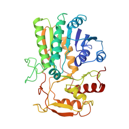
Crystal structures of the oxidized and reduced forms of UDP-galactose 4-epimerase isolated from Escherichia coli.

Thoden, J.B.Frey, P.A.Holden, H.M.

(1996) Biochemistry 35: 2557-2566

  • DOI: https://doi.org/10.1021/bi952715y
  • Primary Citation Related Structures: 
    1NAH, 1NAI

  • PubMed Abstract: 

    UDP-galactose 4-epimerase catalyzes the conversion of UDP-galactose to UDP-glucose through a mechanism involving the transient reduction of NAD+. Here we describe the X-ray structures for epimerase complexed with NADH/UDP, and NAD+/UDP, refined to 1.8 and 2.0 angstrom, respectively. The alpha-carbon positions for the two forms of the enzyme are superimposed with a root-mean-square deviation of 0.36 A. Overall, the models for the reduced and oxidized proteins are very similar except for the positions of several side chains including Phe 178 and Phe 218. The most striking difference between the oxidized and reduced enzymes is the conformation of the nicotinamide ring of the dinucleotide. In the reduced protein, the nicotinamide ring adopts the anti conformation while in the oxidized enzyme the syn conformation is observed. There are also significant structural differences in UDP binding between the oxidized and reduced forms of the protein which most likely explain the observation that uridine nucleotides bind more tightly to epimerase/NADH than to epimerase/NAD+. Both van der Waals and electrostatic interactions between epimerase and NAD+ are extensive with 35 contacts below 3.2 angstrom as would be expected for enzyme that binds the dinucleotide irreversibly. This is in sharp contrast to the patterns typically observed for the NAD+-dependent dehydrogenases which bind nucleotides in a reversible fashion. While it has been postulated that the active site of epimerase must contain a base, the only potential candidates within approximately 5 A of both the NAD+ and the UDP are Asp 31, Asp 58, and ASP 295. These amino acid residues, however, are intimately involved in nucleotide binding and most likely do not play a role in the actual catalytic mechanism. Thus it may be speculated that an amino acid residue, other than glutamate, aspartate, or histidine, may be functioning as the active site base.


  • Organizational Affiliation
    • Institute for Enzyme Research, University of Wisconsin, Madison, Wisconsin 53705 USA.

Macromolecule Content 

  • Total Structure Weight: 38.58 kDa 
  • Atom Count: 3,199 
  • Modeled Residue Count: 338 
  • Deposited Residue Count: 338 
  • Unique protein chains: 1

Macromolecules

Find similar proteins by:|  3D Structure
Entity ID: 1
MoleculeChains  Sequence LengthOrganismDetailsImage
UDP-GALACTOSE 4-EPIMERASE338Escherichia coliMutation(s): 0 
EC: 5.1.3.2
UniProt
Find proteins for P09147 (Escherichia coli (strain K12))
Explore P09147 
Go to UniProtKB:  P09147
Entity Groups
Sequence Clusters30% Identity50% Identity70% Identity90% Identity95% Identity100% Identity
UniProt GroupP09147
Sequence Annotations
Expand
Reference Sequence

Small Molecules

Ligands 5 Unique
IDChains Name / Formula / InChI Key2D Diagram3D Interactions
NAD

Query on NAD


Download:Ideal Coordinates CCD File
D [auth A]NICOTINAMIDE-ADENINE-DINUCLEOTIDE
C21 H27 N7 O14 P2
BAWFJGJZGIEFAR-NNYOXOHSSA-N
UDP

Query on UDP


Download:Ideal Coordinates CCD File
E [auth A]URIDINE-5'-DIPHOSPHATE
C9 H14 N2 O12 P2
XCCTYIAWTASOJW-XVFCMESISA-N
PEG

Query on PEG


Download:Ideal Coordinates CCD File
G [auth A]DI(HYDROXYETHYL)ETHER
C4 H10 O3
MTHSVFCYNBDYFN-UHFFFAOYSA-N
EDO

Query on EDO


Download:Ideal Coordinates CCD File
F [auth A]1,2-ETHANEDIOL
C2 H6 O2
LYCAIKOWRPUZTN-UHFFFAOYSA-N
NA

Query on NA


Download:Ideal Coordinates CCD File
B [auth A],
C [auth A]
SODIUM ION
Na
FKNQFGJONOIPTF-UHFFFAOYSA-N

Experimental Data & Validation

Experimental Data

  • Method: X-RAY DIFFRACTION
  • Resolution: 1.80 Å
  • R-Value Work:  0.165 (Depositor) 
Space Group: P 32 2 1
Unit Cell:
Length ( Å )Angle ( ˚ )
a = 83.5α = 90
b = 83.5β = 90
c = 108.4γ = 120
Software Package:
Software NamePurpose
TNTrefinement

Structure Validation

View Full Validation Report


Entry History 

Deposition Data

Revision History  (Full details and data files)

  • Version 1.0: 1996-12-23
    Type: Initial release
  • Version 1.1: 2008-03-03
    Changes: Version format compliance
  • Version 1.2: 2011-07-13
    Changes: Derived calculations, Version format compliance
  • Version 1.3: 2011-11-16
    Changes: Atomic model
  • Version 1.4: 2017-11-29
    Changes: Derived calculations, Other
  • Version 2.0: 2024-02-14
    Changes: Atomic model, Data collection, Database references, Derived calculations