1NA0 | pdb_00001na0

Design of Stable alpha-Helical Arrays from an Idealized TPR Motif


Experimental Data Snapshot

  • Method: X-RAY DIFFRACTION
  • Resolution: 1.60 Å
  • R-Value Free: 
    0.216 (Depositor) 
  • R-Value Work: 
    0.183 (Depositor) 
  • R-Value Observed: 
    0.186 (Depositor) 

wwPDB Validation 3D Report Full Report

Validation slider image for 1NA0

This is version 1.4 of the entry. See complete history

Literature

Design of Stable alpha-Helical Arrays from an Idealized TPR Motif

Main, E.Xiong, Y.Cocco, M.D'Andrea, L.Regan, L.

(2003) Structure 11: 497-508

  • DOI: https://doi.org/10.1016/s0969-2126(03)00076-5
  • Primary Citation Related Structures: 
    1NA0, 1NA3

  • PubMed Abstract: 

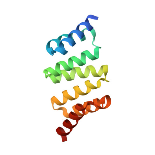
    The tetratricopeptide repeat (TPR) is a 34-amino acid alpha-helical motif that occurs in over 300 different proteins. In the different proteins, three to sixteen or more TPR motifs occur in tandem arrays and function to mediate protein-protein interactions. The binding specificity of each TPR protein is different, although the underlying structural motif is the same. Here we describe a statistical approach to the design of an idealized TPR motif. We present the high-resolution X-ray crystal structures (to 1.55 and 1.6 A) of designed TPR proteins and describe their solution properties and stability. A detailed analysis of these structures provides an understanding of the TPR motif, how it is repeated to give helical arrays with different superhelical twists, and how a very stable framework may be constructed for future functional designs.


  • Organizational Affiliation
    • Department of Molecular Biophysics and Biochemistry, Yale University, New Haven, CT 06520, USA.

Macromolecule Content 

  • Total Structure Weight: 30.42 kDa 
  • Atom Count: 2,166 
  • Modeled Residue Count: 238 
  • Deposited Residue Count: 250 
  • Unique protein chains: 1

Macromolecules

Find similar proteins by:|  3D Structure
Entity ID: 1
MoleculeChains  Sequence LengthOrganismDetailsImage
designed protein CTPR3
A, B
125unidentifiedMutation(s): 0 

Small Molecules

Ligands 6 Unique
IDChains Name / Formula / InChI Key2D Diagram3D Interactions
IPT

Query on IPT


Download:Ideal Coordinates CCD File
M [auth B],
N [auth B]
1-methylethyl 1-thio-beta-D-galactopyranoside
C9 H18 O5 S
BPHPUYQFMNQIOC-NXRLNHOXSA-N
PB

Query on PB


Download:Ideal Coordinates CCD File
C [auth A],
D [auth A],
F [auth B],
G [auth B],
H [auth B]
LEAD (II) ION
Pb
RVPVRDXYQKGNMQ-UHFFFAOYSA-N
ACT

Query on ACT


Download:Ideal Coordinates CCD File
E [auth B]ACETATE ION
C2 H3 O2
QTBSBXVTEAMEQO-UHFFFAOYSA-M
CL

Query on CL


Download:Ideal Coordinates CCD File
I [auth B]CHLORIDE ION
Cl
VEXZGXHMUGYJMC-UHFFFAOYSA-M
MG

Query on MG


Download:Ideal Coordinates CCD File
K [auth B]MAGNESIUM ION
Mg
JLVVSXFLKOJNIY-UHFFFAOYSA-N
NA

Query on NA


Download:Ideal Coordinates CCD File
J [auth B],
L [auth B]
SODIUM ION
Na
FKNQFGJONOIPTF-UHFFFAOYSA-N

Experimental Data & Validation

Experimental Data

  • Method: X-RAY DIFFRACTION
  • Resolution: 1.60 Å
  • R-Value Free:  0.216 (Depositor) 
  • R-Value Work:  0.183 (Depositor) 
  • R-Value Observed: 0.186 (Depositor) 
Space Group: C 1 2 1
Unit Cell:
Length ( Å )Angle ( ˚ )
a = 102.734α = 90
b = 46.56β = 99.11
c = 52.517γ = 90
Software Package:
Software NamePurpose
HKL-2000data collection
SCALEPACKdata scaling
AMoREphasing
REFMACrefinement
HKL-2000data reduction

Structure Validation

View Full Validation Report


Entry History 

Deposition Data

Revision History  (Full details and data files)

  • Version 1.0: 2003-06-03
    Type: Initial release
  • Version 1.1: 2008-04-28
    Changes: Version format compliance
  • Version 1.2: 2011-07-13
    Changes: Version format compliance
  • Version 1.3: 2020-07-29
    Type: Remediation
    Reason: Carbohydrate remediation
    Changes: Data collection, Derived calculations, Structure summary
  • Version 1.4: 2024-02-14
    Changes: Data collection, Database references, Structure summary