1N9G | pdb_00001n9g

Mitochondrial 2-enoyl thioester reductase Etr1p/Etr2p heterodimer from Candida tropicalis


Experimental Data Snapshot

  • Method: X-RAY DIFFRACTION
  • Resolution: 1.98 Å
  • R-Value Free: 
    0.235 (Depositor), 0.280 (DCC) 
  • R-Value Work: 
    0.195 (Depositor), 0.257 (DCC) 
  • R-Value Observed: 
    0.197 (Depositor) 

Starting Model: experimental
View more details

wwPDB Validation 3D Report Full Report

Validation slider image for 1N9G

Ligand Structure Quality Assessment 


This is version 1.5 of the entry. See complete history

Literature

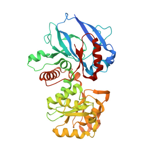
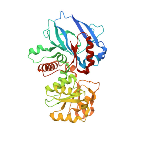
Candida tropicalis expresses two mitochondrial 2-enoyl thioester reductases that are able to form both homodimers and heterodimers.

Torkko, J.M.Koivuranta, K.T.Kastaniotis, A.J.Airenne, T.T.Glumoff, T.Ilves, M.Hartig, A.Gurvitz, A.Hiltunen, J.K.

(2003) J Biological Chem 278: 41213-41220

  • DOI: https://doi.org/10.1074/jbc.M307664200
  • Primary Citation Related Structures: 
    1N9G

  • PubMed Abstract: 

    Here we report on the cloning of a Candida tropicalis gene, ETR2, that is closely related to ETR1. Both genes encode enzymatically active 2-enoyl thioester reductases involved in mitochondrial synthesis of fatty acids (fatty acid synthesis type II) and respiratory competence. The 5'- and 3'-flanking (coding) regions of ETR2 and ETR1 are about 90% (97%) identical, indicating that the genes have evolved via gene duplication. The gene products differ in three amino acid residues: Ile67 (Val), Ala92 (Thr), and Lys251 (Arg) in Etr2p (Etr1p). Quantitative PCR analysis and reverse transcriptase-PCR indicated that both genes were expressed about equally in fermenting and ETR1 predominantly respiring yeast cells. Like the situation with ETR1, expression of ETR2 in respiration-deficient Saccharomyces cerevisiae mutant cells devoid of Ybr026p/Etr1p was able to restore growth on glycerol. Triclosan that is used as an antibacterial agent against fatty acid synthesis type II 2-enoyl thioester reductases inhibited growth of FabI overexpressing mutant yeast cells but was not able to inhibit respiratory growth of the ETR2- or ETR1-complemented mutant yeast cells. Resolving of crystal structures obtained via Etr2p and Etr1p co-crystallization indicated that all possible dimer variants occur in the same asymmetric unit, suggesting that similar dimer formation also takes place in vivo.


  • Organizational Affiliation
    • Biocenter Oulu, Department of Biochemistry, University of Oulu, Finland.

Macromolecule Content 

  • Total Structure Weight: 255.99 kDa 
  • Atom Count: 18,417 
  • Modeled Residue Count: 2,184 
  • Deposited Residue Count: 2,316 
  • Unique protein chains: 2

Macromolecules

Find similar proteins by:|  3D Structure
Entity ID: 1
MoleculeChains  Sequence LengthOrganismDetailsImage
2,4-dienoyl-CoA reductase
A, C, F
386Candida tropicalisMutation(s): 0 
Gene Names: ETR2
EC: 1.3.1.104
UniProt
Find proteins for Q8WZM4 (Candida tropicalis)
Explore Q8WZM4 
Go to UniProtKB:  Q8WZM4
Entity Groups
Sequence Clusters30% Identity50% Identity70% Identity90% Identity95% Identity100% Identity
UniProt GroupQ8WZM4
Sequence Annotations
Expand
Reference Sequence
Find similar proteins by:|  3D Structure
Entity ID: 2
MoleculeChains  Sequence LengthOrganismDetailsImage
2,4-dienoyl-CoA reductase
B, D, E
386Candida tropicalisMutation(s): 0 
Gene Names: ETR1
EC: 1.3.1.104
UniProt
Find proteins for Q8WZM3 (Candida tropicalis)
Explore Q8WZM3 
Go to UniProtKB:  Q8WZM3
Entity Groups
Sequence Clusters30% Identity50% Identity70% Identity90% Identity95% Identity100% Identity
UniProt GroupQ8WZM3
Sequence Annotations
Expand
Reference Sequence

Small Molecules

Ligands 2 Unique
IDChains Name / Formula / InChI Key2D Diagram3D Interactions
NAP

Query on NAP


Download:Ideal Coordinates CCD File
H [auth A],
J [auth B],
O [auth E]
NADP NICOTINAMIDE-ADENINE-DINUCLEOTIDE PHOSPHATE
C21 H28 N7 O17 P3
XJLXINKUBYWONI-NNYOXOHSSA-N
SO4

Query on SO4


Download:Ideal Coordinates CCD File
G [auth A]
I [auth B]
K [auth C]
L [auth D]
M [auth D]
G [auth A],
I [auth B],
K [auth C],
L [auth D],
M [auth D],
N [auth E],
P [auth F]
SULFATE ION
O4 S
QAOWNCQODCNURD-UHFFFAOYSA-L

Experimental Data & Validation

Experimental Data

  • Method: X-RAY DIFFRACTION
  • Resolution: 1.98 Å
  • R-Value Free:  0.235 (Depositor), 0.280 (DCC) 
  • R-Value Work:  0.195 (Depositor), 0.257 (DCC) 
  • R-Value Observed: 0.197 (Depositor) 
Space Group: C 1 2 1
Unit Cell:
Length ( Å )Angle ( ˚ )
a = 229.453α = 90
b = 95.501β = 124.71
c = 164.229γ = 90
Software Package:
Software NamePurpose
DENZOdata reduction
SCALEPACKdata scaling
AMoREphasing
REFMACrefinement

Structure Validation

View Full Validation Report


Ligand Structure Quality Assessment 


Entry History 

Deposition Data

Revision History  (Full details and data files)

  • Version 1.0: 2003-12-09
    Type: Initial release
  • Version 1.1: 2008-04-28
    Changes: Version format compliance
  • Version 1.2: 2011-07-13
    Changes: Version format compliance
  • Version 1.3: 2018-03-07
    Changes: Advisory, Data collection
  • Version 1.4: 2023-08-16
    Changes: Advisory, Data collection, Database references, Derived calculations, Refinement description
  • Version 1.5: 2024-12-25
    Changes: Derived calculations, Structure summary