1N8S | pdb_00001n8s

Structure of the pancreatic lipase-colipase complex


Experimental Data Snapshot

  • Method: X-RAY DIFFRACTION
  • Resolution: 3.04 Å
  • R-Value Free: 
    0.290 (Depositor) 
  • R-Value Work: 
    0.230 (Depositor) 
  • R-Value Observed: 
    0.230 (Depositor) 

wwPDB Validation 3D Report Full Report

Validation slider image for 1N8S

This is version 1.4 of the entry. See complete history

Literature

Structure of the pancreatic lipase-procolipase complex

van Tilbeurgh, H.Sarda, L.Verger, R.Cambillau, C.

(1992) Nature 359: 159-162

  • DOI: https://doi.org/10.1038/359159a0
  • Primary Citation Related Structures: 
    1N8S

  • PubMed Abstract: 

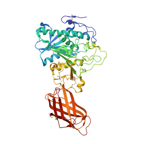
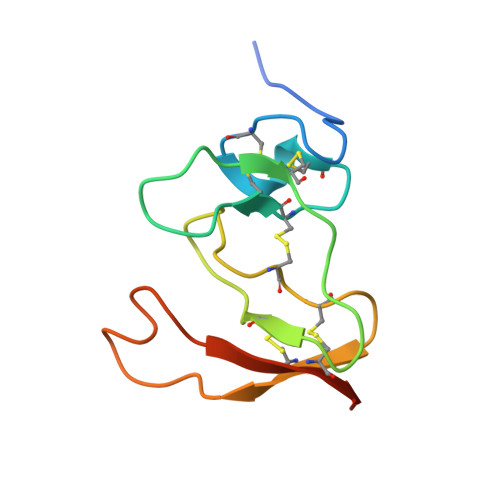
    Interfacial adsorption of pancreatic lipase is strongly dependent on the physical chemical properties of the lipid surface. These properties are affected by amphiphiles such as phospholipids and bile salts. In the presence of such amphiphiles, lipase binding to the interface requires a protein cofactor, colipase. We obtained crystals of the pancreatic lipase-procolipase complex and solved the structure at 3.04 A resolution. Here we describe the structure of procolipase, which essentially consists of three 'fingers' and is topologically comparable to snake toxins. The tips of the fingers contain most of the hydrophobic amino acids and presumably form the interfacial binding site. Lipase binding occurs at the opposite side to this site and involves polar interactions. Determination of the three-dimensional structure of pancreatic lipase has revealed the presence of two domains: an amino-terminal domain, at residues 1-336 containing the active site and a carboxy-terminal domain at residues 337-449 (ref. 6). Procolipase binds exclusively to the C-terminal domain of lipase. No conformational change in the lipase molecule is induced by the binding of procolipase.


  • Organizational Affiliation
    • LCCMB-CNRS, Faculté de Médecine Nord, Marseille, France.

Macromolecule Content 

  • Total Structure Weight: 59.92 kDa 
  • Atom Count: 4,131 
  • Modeled Residue Count: 534 
  • Deposited Residue Count: 544 
  • Unique protein chains: 2

Macromolecules

Find similar proteins by:|  3D Structure
Entity ID: 1
MoleculeChains  Sequence LengthOrganismDetailsImage
Triacylglycerol lipase, pancreatic449Homo sapiensMutation(s): 0 
EC: 3.1.1.3
Membrane Entity: Yes 
UniProt & NIH Common Fund Data Resources
Find proteins for P16233 (Homo sapiens)
Explore P16233 
Go to UniProtKB:  P16233
PHAROS:  P16233
GTEx:  ENSG00000175535 
Entity Groups
Sequence Clusters30% Identity50% Identity70% Identity90% Identity95% Identity100% Identity
UniProt GroupP16233
Sequence Annotations
Expand
Reference Sequence
Find similar proteins by:|  3D Structure
Entity ID: 2
MoleculeChains  Sequence LengthOrganismDetailsImage
colipase IIB [auth C]95Sus scrofaMutation(s): 0 
Membrane Entity: Yes 
UniProt
Find proteins for P02703 (Sus scrofa)
Explore P02703 
Go to UniProtKB:  P02703
Entity Groups
Sequence Clusters30% Identity50% Identity70% Identity90% Identity95% Identity100% Identity
UniProt GroupP02703
Sequence Annotations
Expand
Reference Sequence

Experimental Data & Validation

Experimental Data

  • Method: X-RAY DIFFRACTION
  • Resolution: 3.04 Å
  • R-Value Free:  0.290 (Depositor) 
  • R-Value Work:  0.230 (Depositor) 
  • R-Value Observed: 0.230 (Depositor) 
Space Group: P 32 2 1
Unit Cell:
Length ( Å )Angle ( ˚ )
a = 80.3α = 90
b = 80.3β = 90
c = 251γ = 120
Software Package:
Software NamePurpose
XDSdata scaling
AUTOMARdata reduction
MLPHAREphasing
X-PLORrefinement
XDSdata reduction

Structure Validation

View Full Validation Report


Entry History 

Deposition Data

Revision History  (Full details and data files)

  • Version 1.0: 2002-12-18
    Type: Initial release
  • Version 1.1: 2008-04-28
    Changes: Version format compliance
  • Version 1.2: 2011-07-13
    Changes: Version format compliance
  • Version 1.3: 2017-10-11
    Changes: Refinement description
  • Version 1.4: 2024-11-06
    Changes: Data collection, Database references, Structure summary