1N80

Bacteriophage T4 baseplate structural protein gp8


Experimental Data Snapshot

  • Method: X-RAY DIFFRACTION
  • Resolution: 2.45 Å
  • R-Value Free: 0.284 
  • R-Value Work: 0.236 
  • R-Value Observed: 0.236 

wwPDB Validation   3D Report Full Report


This is version 1.3 of the entry. See complete history


Literature

Structure and location of gene product 8 in the bacteriophage T4 baseplate

Leiman, P.G.Shneider, M.M.Kostyuchenko, V.A.Chipman, P.R.Mesyanzhinov, V.V.Rossmann, M.G.

(2003) J Mol Biol 328: 821-833

  • DOI: https://doi.org/10.1016/s0022-2836(03)00366-8
  • Primary Citation of Related Structures:  
    1N7Z, 1N80, 1N8B

  • PubMed Abstract: 

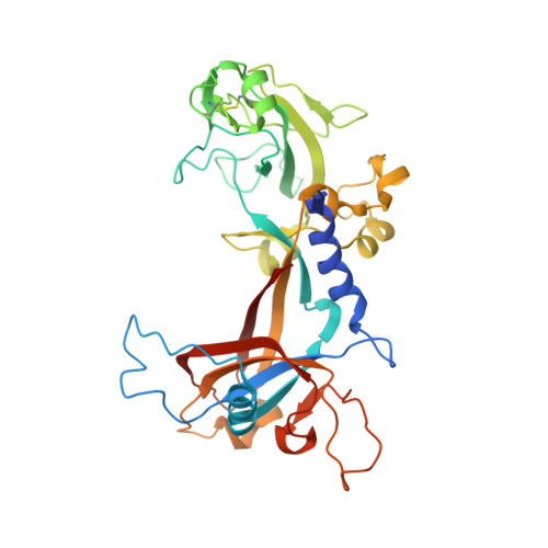
    Many bacteriophages, such as T4, T7, RB49, and phi29, have complex, sometimes multilayered, tails that facilitate an almost 100% success rate for the viral particles to infect host cells. In bacteriophage T4, there is a baseplate, which is a multiprotein assembly, at the distal end of the contractile tail. The baseplate communicates to the tail that the phage fibers have attached to the host cell, thereby initiating the infection process. Gene product 8 (gp8), whose amino acid sequence consists of 334 residues, is one of at least 16 different structural proteins that constitute the T4 baseplate and is the sixth baseplate protein whose structure has been determined. A 2.0A resolution X-ray structure of gp8 shows that the two-domain protein forms a dimer, in which each monomer consists of a three-layered beta-sandwich with two loops, each containing an alpha-helix at the opposite sides of the sandwich. The crystals of gp8 were produced in the presence of concentrated chloride and bromide ions, resulting in at least 11 halide-binding sites per monomer. Five halide sites, situated at the N termini of alpha-helices, have a protein environment observed in other halide-containing protein crystal structures. The computer programs EMfit and SITUS were used to determine the positions of six gp8 dimers within the 12A resolution cryo-electron microscopy image reconstruction of the baseplate-tail tube complex. The gp8 dimers were found to be located in the upper part of the baseplate outer rim. About 20% of the gp8 surface is involved in contacts with other baseplate proteins, presumed to be gp6, gp7, and gp10. With the structure determination of gp8, a total of 53% of the volume of the baseplate has now been interpreted in terms of its atomic structure.


  • Organizational Affiliation

    Department of Biological Sciences, Purdue University, Lilly Hall of Life Sciences, West Lafayette, IN 47907-2054, USA.


Macromolecules
Find similar proteins by:  (by identity cutoff)  |  3D Structure
Entity ID: 1
MoleculeChains Sequence LengthOrganismDetailsImage
baseplate structural protein gp8
A, B, C, D
334Tequatrovirus T4Mutation(s): 0 
Gene Names: gene 8
UniProt
Find proteins for P19062 (Enterobacteria phage T4)
Explore P19062 
Go to UniProtKB:  P19062
Entity Groups  
Sequence Clusters30% Identity50% Identity70% Identity90% Identity95% Identity100% Identity
UniProt GroupP19062
Sequence Annotations
Expand
  • Reference Sequence
Small Molecules
Ligands 1 Unique
IDChains Name / Formula / InChI Key2D Diagram3D Interactions
CL
Query on CL

Download Ideal Coordinates CCD File 
AA [auth C]
BA [auth C]
CA [auth D]
DA [auth D]
E [auth A]
AA [auth C],
BA [auth C],
CA [auth D],
DA [auth D],
E [auth A],
EA [auth D],
F [auth A],
FA [auth D],
G [auth A],
GA [auth D],
H [auth A],
HA [auth D],
I [auth A],
IA [auth D],
J [auth A],
JA [auth D],
K [auth A],
L [auth A],
M [auth A],
N [auth A],
O [auth B],
P [auth B],
Q [auth B],
R [auth B],
S [auth B],
T [auth B],
U [auth C],
V [auth C],
W [auth C],
X [auth C],
Y [auth C],
Z [auth C]
CHLORIDE ION
Cl
VEXZGXHMUGYJMC-UHFFFAOYSA-M
Experimental Data & Validation

Experimental Data

  • Method: X-RAY DIFFRACTION
  • Resolution: 2.45 Å
  • R-Value Free: 0.284 
  • R-Value Work: 0.236 
  • R-Value Observed: 0.236 
  • Space Group: P 1
Unit Cell:
Length ( Å )Angle ( ˚ )
a = 66.869α = 93.11
b = 66.777β = 101.28
c = 82.158γ = 90.77
Software Package:
Software NamePurpose
DENZOdata reduction
SCALEPACKdata scaling
AMoREphasing
CNSrefinement

Structure Validation

View Full Validation Report


Entry History 

Deposition Data

Revision History  (Full details and data files)

  • Version 1.0: 2003-06-10
    Type: Initial release
  • Version 1.1: 2008-04-28
    Changes: Version format compliance
  • Version 1.2: 2011-07-13
    Changes: Version format compliance
  • Version 1.3: 2023-08-16
    Changes: Data collection, Database references, Derived calculations, Refinement description