1N69 | pdb_00001n69

Crystal structure of human saposin B


Experimental Data Snapshot

  • Method: X-RAY DIFFRACTION
  • Resolution: 2.20 Å
  • R-Value Free: 
    0.262 (Depositor), 0.265 (DCC) 
  • R-Value Work: 
    0.222 (Depositor), 0.221 (DCC) 
  • R-Value Observed: 
    0.222 (Depositor) 

wwPDB Validation 3D Report Full Report

Validation slider image for 1N69

Ligand Structure Quality Assessment 


This is version 2.1 of the entry. See complete history

Literature

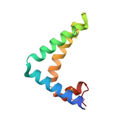
Crystal Structure of saposin B reveals a dimeric shell for lipid binding

Ahn, V.E.Faull, K.F.Whitelegge, J.P.Fluharty, A.L.Prive, G.G.

(2003) Proc Natl Acad Sci U S A 100: 38-43

  • DOI: https://doi.org/10.1073/pnas.0136947100
  • Primary Citation Related Structures: 
    1N69

  • PubMed Abstract: 

    Saposin B is a small, nonenzymatic glycosphingolipid activator protein required for the breakdown of cerebroside sulfates (sulfatides) within the lysosome. The protein can extract target lipids from membranes, forming soluble protein-lipid complexes that are recognized by arylsulfatase A. The crystal structure of human saposin B reveals an unusual shell-like dimer consisting of a monolayer of alpha-helices enclosing a large hydrophobic cavity. Although the secondary structure of saposin B is similar to that of the known monomeric members of the saposin-like superfamily, the helices are repacked into a different tertiary arrangement to form the homodimer. A comparison of the two forms of the saposin B dimer suggests that extraction of target lipids from membranes involves a conformational change that facilitates access to the inner cavity.


  • Organizational Affiliation
    • Department of Medical Biophysics, University of Toronto, ON, Canada M5G 2M9.

Macromolecule Content 

  • Total Structure Weight: 28.13 kDa 
  • Atom Count: 1,955 
  • Modeled Residue Count: 236 
  • Deposited Residue Count: 243 
  • Unique protein chains: 1

Macromolecules

Find similar proteins by:|  3D Structure
Entity ID: 1
MoleculeChains  Sequence LengthOrganismDetailsImage
SAPOSIN B
A, B, C
81Homo sapiensMutation(s): 0 
Gene Names: ProsaposinPSAP
Membrane Entity: Yes 
UniProt & NIH Common Fund Data Resources
Find proteins for P07602 (Homo sapiens)
Explore P07602 
Go to UniProtKB:  P07602
PHAROS:  P07602
GTEx:  ENSG00000197746 
Entity Groups
Sequence Clusters30% Identity50% Identity70% Identity90% Identity95% Identity100% Identity
UniProt GroupP07602
Sequence Annotations
Expand
Reference Sequence

Small Molecules

Ligands 1 Unique
IDChains Name / Formula / InChI Key2D Diagram3D Interactions
3PE

Query on 3PE


Download:Ideal Coordinates CCD File
D [auth A]1,2-Distearoyl-sn-glycerophosphoethanolamine
C41 H82 N O8 P
LVNGJLRDBYCPGB-LDLOPFEMSA-N

Experimental Data & Validation

Experimental Data

  • Method: X-RAY DIFFRACTION
  • Resolution: 2.20 Å
  • R-Value Free:  0.262 (Depositor), 0.265 (DCC) 
  • R-Value Work:  0.222 (Depositor), 0.221 (DCC) 
  • R-Value Observed: 0.222 (Depositor) 
Space Group: P 31 2 1
Unit Cell:
Length ( Å )Angle ( ˚ )
a = 72.14α = 90
b = 72.14β = 90
c = 94.366γ = 120
Software Package:
Software NamePurpose
MOSFLMdata reduction
SCALAdata scaling
SnBphasing
SHARPphasing
DMmodel building
CNSrefinement
CCP4data scaling

Structure Validation

View Full Validation Report


Ligand Structure Quality Assessment 


Entry History 

Deposition Data

Revision History  (Full details and data files)

  • Version 1.0: 2003-01-07
    Type: Initial release
  • Version 1.1: 2008-04-28
    Changes: Version format compliance
  • Version 1.2: 2011-07-13
    Changes: Version format compliance
  • Version 1.3: 2017-10-11
    Changes: Refinement description
  • Version 2.0: 2021-06-30
    Changes: Advisory, Atomic model, Data collection, Database references, Derived calculations, Non-polymer description, Structure summary
  • Version 2.1: 2024-10-30
    Changes: Data collection, Database references, Structure summary