1N0Y | pdb_00001n0y

Crystal Structure of Pb-bound Calmodulin


Experimental Data Snapshot

  • Method: X-RAY DIFFRACTION
  • Resolution: 1.75 Å
  • R-Value Free: 
    0.223 (Depositor), 0.219 (DCC) 
  • R-Value Work: 
    0.213 (Depositor) 
  • R-Value Observed: 
    0.214 (Depositor) 

wwPDB Validation 3D Report Full Report

Validation slider image for 1N0Y

This is version 1.4 of the entry. See complete history

Literature

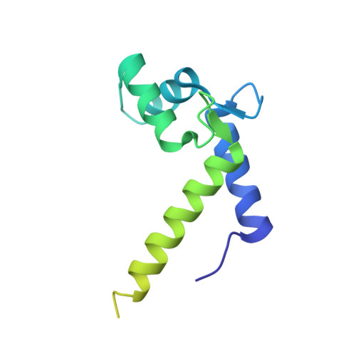
Domain flexibility in the 1.75 A resolution structure of Pb2+-calmodulin.

Wilson, M.A.Brunger, A.T.

(2003) Acta Crystallogr D Biol Crystallogr 59: 1782-1792

  • DOI: https://doi.org/10.1107/s0907444903016846
  • Primary Citation Related Structures: 
    1N0Y

  • PubMed Abstract: 

    Calmodulin (CaM) regulates a variety of cellular processes by interacting with a large number of proteins in a Ca(2+)-dependent manner. Conformational flexibility plays a key role in CaM function, although the full extent and detailed features of this flexibility are not fully characterized. Here, the 1.75 A resolution crystal structure of Pb(2+)-bound Paramecium tetraurelia CaM crystallized in a previously unobserved monoclinic lattice is reported. Pb(2+)-CaM is disordered in this new lattice and only a portion of each of the two molecules in the asymmetric unit can be modeled. Comparison of the structures of Ca(2+)-CaM and Pb(2+)-CaM show close agreement in the C-terminal domain but significant structural differences in the N-terminal domain. In addition, translation-libration-screw (TLS) refinement and Rosenfield difference analysis reveal inter-helical flexibility in the metal-bound N-terminal domain of the protein that is absent in the metal-bound C-terminal domain and indicates that the two structurally similar domains of CaM are dynamically distinct. These results demonstrate that TLS refinement and Rosenfield difference analysis allow detailed information about macromolecular flexibility to be extracted from X-ray diffraction data even when the crystal lattice prohibits full manifestation of this flexibility.


  • Organizational Affiliation
    • Rosenstiel Basic Medical Sciences Research Center, Brandeis University, 415 South Street, Waltham, MA 02453, USA.

Macromolecule Content 

  • Total Structure Weight: 36.47 kDa 
  • Atom Count: 1,483 
  • Modeled Residue Count: 166 
  • Deposited Residue Count: 296 
  • Unique protein chains: 1

Macromolecules

Find similar proteins by:|  3D Structure
Entity ID: 1
MoleculeChains  Sequence LengthOrganismDetailsImage
Calmodulin
A, B
148Paramecium tetraureliaMutation(s): 0 
Gene Names: CAM
UniProt
Find proteins for P07463 (Paramecium tetraurelia)
Explore P07463 
Go to UniProtKB:  P07463
Entity Groups
Sequence Clusters30% Identity50% Identity70% Identity90% Identity95% Identity100% Identity
UniProt GroupP07463
Sequence Annotations
Expand
Reference Sequence

Small Molecules

Ligands 3 Unique
IDChains Name / Formula / InChI Key2D Diagram3D Interactions
PB

Query on PB


Download:Ideal Coordinates CCD File
C [auth A]
D [auth A]
E [auth A]
F [auth A]
G [auth A]
C [auth A],
D [auth A],
E [auth A],
F [auth A],
G [auth A],
H [auth A],
I [auth A],
K [auth B],
L [auth B],
M [auth B],
N [auth B],
O [auth B],
P [auth B],
Q [auth B]
LEAD (II) ION
Pb
RVPVRDXYQKGNMQ-UHFFFAOYSA-N
CAC

Query on CAC


Download:Ideal Coordinates CCD File
J [auth A]CACODYLATE ION
C2 H6 As O2
OGGXGZAMXPVRFZ-UHFFFAOYSA-M
ACT

Query on ACT


Download:Ideal Coordinates CCD File
R [auth B]ACETATE ION
C2 H3 O2
QTBSBXVTEAMEQO-UHFFFAOYSA-M

Experimental Data & Validation

Experimental Data

  • Method: X-RAY DIFFRACTION
  • Resolution: 1.75 Å
  • R-Value Free:  0.223 (Depositor), 0.219 (DCC) 
  • R-Value Work:  0.213 (Depositor) 
  • R-Value Observed: 0.214 (Depositor) 
Space Group: C 1 2 1
Unit Cell:
Length ( Å )Angle ( ˚ )
a = 100.936α = 90
b = 30.792β = 109.31
c = 113.084γ = 90
Software Package:
Software NamePurpose
DENZOdata reduction
SCALEPACKdata scaling
SHELXDphasing
SHARPphasing
REFMACrefinement

Structure Validation

View Full Validation Report


Entry History 

Deposition Data

Revision History  (Full details and data files)

  • Version 1.0: 2003-09-30
    Type: Initial release
  • Version 1.1: 2008-04-28
    Changes: Version format compliance
  • Version 1.2: 2011-07-13
    Changes: Non-polymer description, Version format compliance
  • Version 1.3: 2017-10-11
    Changes: Refinement description
  • Version 1.4: 2024-02-14
    Changes: Data collection, Database references, Derived calculations