1MXS

Crystal structure of 2-keto-3-deoxy-6-phosphogluconate (KDPG) aldolase from Pseudomonas putida.


Experimental Data Snapshot

  • Method: X-RAY DIFFRACTION
  • Resolution: 2.20 Å
  • R-Value Free: 0.214 
  • R-Value Work: 0.171 
  • R-Value Observed: 0.171 

wwPDB Validation   3D Report Full Report


This is version 1.4 of the entry. See complete history


Literature

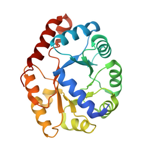
Structure of 2-keto-3-deoxy-6-phosphogluconate (KDPG) aldolase from Pseudomonas putida.

Bell, B.J.Watanabe, L.Rios-Steiner, J.L.Tulinsky, A.Lebioda, L.Arni, R.K.

(2003) Acta Crystallogr D Biol Crystallogr 59: 1454-1458

  • DOI: https://doi.org/10.1107/s0907444903013192
  • Primary Citation of Related Structures:  
    1MXS

  • PubMed Abstract: 

    2-Keto-3-deoxy-6-phosphogluconate (KDPG) aldolase from Pseudomonas putida is a key enzyme in the Entner-Doudoroff pathway which catalyses the cleavage of KDPG via a class I Schiff-base mechanism. The crystal structure of this enzyme has been refined to a crystallographic residual R = 17.1% (R(free) = 21.4%). The N-terminal helix caps one side of the torus of the (betaalpha)(8)-barrel and the active site is located on the opposite, carboxylic side of the barrel. The Schiff-base-forming Lys145 is coordinated by a sulfate (or phosphate) ion and two solvent water molecules. The interactions that stabilize the trimer are predominantly hydrophobic, with the exception of the cyclically permuted bonds formed between Glu132 OE1 of one molecule and Thr129 OG1 of a symmetry-equivalent molecule. Except for the N-terminal helix, the structure of KDPG aldolase from P. putida closely resembles the structure of the homologous enzyme from Escherichia coli.


  • Organizational Affiliation

    Department of Chemistry, Michigan State University, East Lansing, MI 48824, USA.


Macromolecules
Find similar proteins by:  (by identity cutoff)  |  3D Structure
Entity ID: 1
MoleculeChains Sequence LengthOrganismDetailsImage
KDPG Aldolase225Pseudomonas putidaMutation(s): 0 
EC: 4.1.2.14
UniProt
Find proteins for P00885 (Pseudomonas putida)
Explore P00885 
Go to UniProtKB:  P00885
Entity Groups  
Sequence Clusters30% Identity50% Identity70% Identity90% Identity95% Identity100% Identity
UniProt GroupP00885
Sequence Annotations
Expand
  • Reference Sequence
Experimental Data & Validation

Experimental Data

  • Method: X-RAY DIFFRACTION
  • Resolution: 2.20 Å
  • R-Value Free: 0.214 
  • R-Value Work: 0.171 
  • R-Value Observed: 0.171 
  • Space Group: P 21 3
Unit Cell:
Length ( Å )Angle ( ˚ )
a = 103.3α = 90
b = 103.3β = 90
c = 103.3γ = 90
Software Package:
Software NamePurpose
SCALEPACKdata scaling
AMoREphasing
CNSrefinement

Structure Validation

View Full Validation Report


Entry History 

Deposition Data

Revision History  (Full details and data files)

  • Version 1.0: 2003-09-16
    Type: Initial release
  • Version 1.1: 2008-04-28
    Changes: Version format compliance
  • Version 1.2: 2011-07-13
    Changes: Derived calculations, Version format compliance
  • Version 1.3: 2017-10-11
    Changes: Refinement description
  • Version 1.4: 2023-10-25
    Changes: Data collection, Database references, Derived calculations, Refinement description