Preliminary crystallographic analysis of manganese peroxidase from Phanerochaete chrysosporium.
Sundaramoorthy, M., Kishi, K., Gold, M.H., Poulos, T.L.(1994) J Mol Biol 238: 845-848
- PubMed: 8182752 Search on PubMed
- DOI: https://doi.org/10.1006/jmbi.1994.1338
- Primary Citation Related Structures:
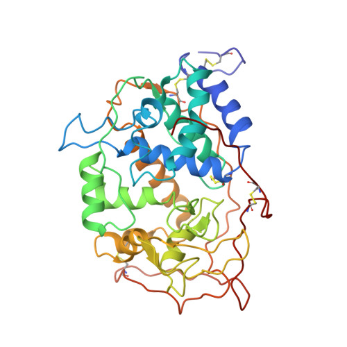
1MNP - PubMed Abstract:
Manganese peroxidase from the white rot basidiomycete Phanerochaete chrysosporium has been crystallized in a form suitable for high-resolution X-ray structure determination. Crystals were grown from solutions containing 30% polyethylene glycol 8000, ammonium sulfate and cacodylate buffer at pH 6.5, using macroseeding techniques. A complete data set has been obtained to 2.06 A resolution. The data can be indexed in space group P1 with a = 45.96 A, b = 53.77 A, c = 84.87 A, alpha = 97.01 degrees, beta = 105.72 degrees and gamma = 90.1 degrees, with two peroxidase molecules per asymmetric unit, or in space group C2 with a = 163.23 A, b = 45.97 A, c = 53.72 A and beta = 97.16 degrees, with only one molecule in the assymetric unit. Lignin peroxidase, which shares about 57% sequence identity with manganese peroxidase, was used as a probe for molecular replacement. Unique rotation and translation solutions have been obtained in space groups P1 and C2. The structure has been partially refined in space group C2 to R = 0.22 for data between 10 and 2.06 A.
- Department of Molecular Biology and Biochemistry, University of California Irvine 92717.
Organizational Affiliation: