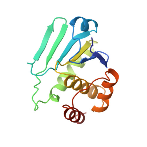
Crystal structure of the MAPK phosphatase Pyst1 catalytic domain and implications for regulated activation.
Stewart, A.E., Dowd, S., Keyse, S.M., McDonald, N.Q.(1999) Nat Struct Biol 6: 174-181
- PubMed: 10048930
- DOI: https://doi.org/10.1038/5861
- Primary Citation of Related Structures:
1MKP - PubMed Abstract:
The crystal structure of the catalytic domain from the MAPK phosphatase Pyst1 (Pyst1-CD) has been determined at 2.35 A. The structure adopts a protein tyrosine phosphatase (PTPase) fold with a shallow active site that displays a distorted geometry in the absence of its substrate with some similarity to the dual-specificity phosphatase cdc25. Functional characterization of Pyst1-CD indicates it is sufficient to dephosphorylate activated ERK2 in vitro. Kinetic analysis of Pyst1 and Pyst1-CD using the substrate p-nitrophenyl phosphate (pNPP) reveals that both molecules undergo catalytic activation in the presence of recombinant inactive ERK2, switching from a low- to high-activity form. Mutation of Asp 262, located 5.5 A distal to the active site, demonstrates it is essential for catalysis in the high-activity ERK2-dependent conformation of Pyst1 but not for the low-activity ERK2-independent form, suggesting that ERK2 induces closure of the Asp 262 loop over the active site, thereby enhancing Pyst1 catalytic efficiency.
- Structural Biology Laboratory, Imperial Cancer Research Fund, London, UK.
Organizational Affiliation:


