1MFG

The Structure of ERBIN PDZ domain bound to the Carboxy-terminal tail of the ErbB2 Receptor


Experimental Data Snapshot

  • Method: X-RAY DIFFRACTION
  • Resolution: 1.25 Å
  • R-Value Free: 0.165 
  • R-Value Work: 0.128 
  • R-Value Observed: 0.130 

wwPDB Validation   3D Report Full Report


This is version 1.2 of the entry. See complete history


Literature

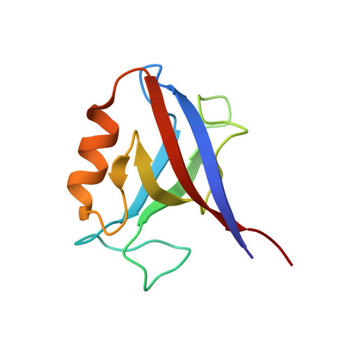
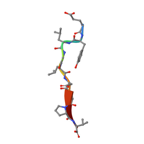
Novel mode of ligand recognition by the erbin PDZ domain

Birrane, G.Chung, J.Ladias, J.A.

(2003) J Biol Chem 278: 1399-1402

  • DOI: https://doi.org/10.1074/jbc.C200571200
  • Primary Citation of Related Structures:  
    1MFG, 1MFL

  • PubMed Abstract: 
  • Erbin contains a class I PDZ domain that binds to the C-terminal region of the receptor tyrosine kinase ErbB2, a class II ligand. The crystal structure of the human Erbin PDZ bound to the peptide EYLGLDVPV corresponding to the C-terminal residues 1247-1255 of human ErbB2 has been determined at 1 ...

    Erbin contains a class I PDZ domain that binds to the C-terminal region of the receptor tyrosine kinase ErbB2, a class II ligand. The crystal structure of the human Erbin PDZ bound to the peptide EYLGLDVPV corresponding to the C-terminal residues 1247-1255 of human ErbB2 has been determined at 1.25-A resolution. The Erbin PDZ deviates from the canonical PDZ fold in that it contains a single alpha-helix. The isopropyl group of valine at position -2 of the ErbB2 peptide interacts with the Erbin Val(1351) and displaces the peptide backbone away from the alpha-helix, elucidating the molecular basis of class II ligand recognition by a class I PDZ domain. Strikingly, the phenolic ring of tyrosine -7 enters into a pocket formed by the extended beta 2-beta 3 loop of the Erbin PDZ. Phosphorylation of tyrosine -7 abolishes this interaction but does not affect the binding of the four C-terminal peptidic residues to PDZ, as revealed by the crystal structure of the Erbin PDZ complexed with a phosphotyrosine-containing ErbB2 peptide. Since phosphorylation of tyrosine -7 plays a critical role in ErbB2 function, the selective binding and sequestration of this residue in its unphosphorylated state by the Erbin PDZ provides a novel mechanism for regulation of the ErbB2-mediated signaling and oncogenicity.


    Related Citations: 
    • BIN: A BASOLATERAL PDZ PROTEIN THAT INTERACTS WITH THE MAMMALIAN ERBB2/HER2 RECEPTOR
      BORG, J.P., MARCHETTO, S., LEBIVIC, A., OLLENDORFF, V., JAULIN-BASTARD, F., SAITO, H., FOURNIER, E., ADELAIDE, J., MARGOLIS, B., BIRNBAUM, D.
      (2000) Nat Cell Biol 2: 407
    • THE ERBIN PDZ DOMAIN BINDS WITH HIGH AFFINITY AND SPECIFICITY TO THE CARBOXYL TERMINI OF DELTA-CATENIN AND ARVCF
      LAURA, R.P., WITT, A.S., HELD, H.A., GERSTNER, R., DESHAYES, K., KOEHLER, M.F., KOSIK, K.S., SIDHU, S.S., LASKY, L.A.
      (2002) J Biol Chem 277: 12906
    • ERBIN IS A PROTEIN CONCENTRATED AT POSTSYNAPTIC MEMBRANES THAT INTERACTS WITH PSD-95
      HUANG, Y.Z., WANG, Q., XIONG, W.C., MEI, L.
      (2001) J Biol Chem 276: 19318
    • THE ERBB2/HER2 RECEPTOR DIFFERENTIALLY INTERACTS WITH ERBIN AND PICK1 PSD-95/DLG/ZO-1 DOMAIN PROTEINS
      JAULIN-BASTARD, F., SAITO, H., LEBIVIC, A., OLLENDORFF, V., MARCHETTO, S., BIRNBAUM, D., BORG, J.P.
      (2001) J Biol Chem 276: 15256

    Organizational Affiliation

    Molecular Medicine Laboratory and Macromolecular Crystallography Unit, Division of Experimental Medicine, Harvard Institutes of Medicine, Harvard Medical School, Boston, Massachusetts 02115, USA.


Macromolecules
Find similar proteins by:  (by identity cutoff)  |  3D Structure
Entity ID: 1
MoleculeChainsSequence LengthOrganismDetailsImage
Erb-B2 INTERACTING PROTEINA95Homo sapiensMutation(s): 0 
Gene Names: ERBINERBB2IPKIAA1225LAP2
UniProt & NIH Common Fund Data Resources
Find proteins for Q96RT1 (Homo sapiens)
Explore Q96RT1 
Go to UniProtKB:  Q96RT1
PHAROS:  Q96RT1
Entity Groups  
Sequence Clusters30% Identity50% Identity70% Identity90% Identity95% Identity100% Identity
UniProt GroupQ96RT1
Protein Feature View
Expand
  • Reference Sequence

Find similar proteins by:  Sequence   |   3D Structure  

Entity ID: 2
MoleculeChainsSequence LengthOrganismDetailsImage
Erb-B2 carboxyl-terminal fragmentB9N/AMutation(s): 0 
UniProt & NIH Common Fund Data Resources
Find proteins for P04626 (Homo sapiens)
Explore P04626 
Go to UniProtKB:  P04626
PHAROS:  P04626
Entity Groups  
UniProt GroupP04626
Protein Feature View
Expand
  • Reference Sequence
Experimental Data & Validation

Experimental Data

  • Method: X-RAY DIFFRACTION
  • Resolution: 1.25 Å
  • R-Value Free: 0.165 
  • R-Value Work: 0.128 
  • R-Value Observed: 0.130 
  • Space Group: P 1 21 1
Unit Cell:
Length ( Å )Angle ( ˚ )
a = 26.605α = 90
b = 57.417β = 100.59
c = 30.439γ = 90
Software Package:
Software NamePurpose
DENZOdata reduction
SCALEPACKdata scaling
SOLVEphasing
SHELXL-97refinement

Structure Validation

View Full Validation Report


Entry History 

Deposition Data

Revision History  (Full details and data files)

  • Version 1.0: 2003-01-21
    Type: Initial release
  • Version 1.1: 2008-04-28
    Changes: Version format compliance
  • Version 1.2: 2011-07-13
    Changes: Version format compliance