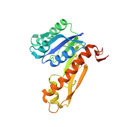
1MEN

complex structure of human GAR Tfase and substrate beta-GAR


Experimental Data Snapshot

  • Method: X-RAY DIFFRACTION
  • Resolution: 2.23 Å
  • R-Value Free: 0.266 
  • R-Value Work: 0.220 

Starting Model: experimental
View more details

wwPDB Validation   3D Report Full Report


This is version 1.4 of the entry. See complete history


Literature

Crystal structures of human GAR Tfase of low and high pH and with substrate beta-GAR

Zhang, Y.Desharnais, J.Greasley, S.E.Beardsley, G.P.Boger, D.L.Wilson, I.A.

(2002) Biochemistry 41: 14206-14215

  • DOI: https://doi.org/10.1021/bi020522m
  • Primary Citation of Related Structures:  
    1MEJ, 1MEN, 1MEO

  • PubMed Abstract: 

    Glycinamide ribonucleotide transformylase (GAR Tfase) is a key folate-dependent enzyme in the de novo purine biosynthesis pathway and, as such, has been the target for antitumor drug design. Here, we describe the crystal structures of the human GAR Tfase (purN) component of the human trifunctional protein (purD-purM-purN) at various pH values and in complex with its substrate. Human GAR Tfase exhibits pH-dependent enzyme activity with its maximum around pH 7.5-8. Comparison of unliganded human GAR Tfase structures at pH 4.2 and pH 8.5 reveals conformational differences in the substrate binding loop, which at pH 4.2 occupies the binding cleft and prohibits substrate binding, while at pH 8.5 is permissive for substrate binding. The crystal structure of GAR Tfase with its natural substrate, beta-glycinamide ribonucleotide (beta-GAR), at pH 8.5 confirms this conformational isomerism. Surprisingly, several important structural differences are found between human GAR Tfase and previously reported E. coli GAR Tfase structures, which have been used as the primary template for drug design studies. While the E. coli structure gave valuable insights into the active site and formyl transfer mechanism, differences in structure and inhibition between the bacterial and mammalian enzymes suggest that the human GAR Tfase structure is now the appropriate template for the design of anti-cancer agents.


  • Organizational Affiliation

    Department of Molecular Biology, The Skaggs Institute for Chemical Biology, The Scripps Research Institute, 10550 North Torrey Pines Road, La Jolla, California 92037, USA.


Macromolecules
Find similar proteins by:  (by identity cutoff)  |  3D Structure
Entity ID: 1
MoleculeChains Sequence LengthOrganismDetailsImage
Phosphoribosylglycinamide formyltransferase
A, B, C
223Homo sapiensMutation(s): 0 
Gene Names: GART
EC: 2.1.2.2
UniProt & NIH Common Fund Data Resources
Find proteins for P22102 (Homo sapiens)
Explore P22102 
Go to UniProtKB:  P22102
PHAROS:  P22102
GTEx:  ENSG00000159131 
Entity Groups  
Sequence Clusters30% Identity50% Identity70% Identity90% Identity95% Identity100% Identity
UniProt GroupP22102
Sequence Annotations
Expand
  • Reference Sequence
Small Molecules
Ligands 1 Unique
IDChains Name / Formula / InChI Key2D Diagram3D Interactions
GAR
Query on GAR

Download Ideal Coordinates CCD File 
D [auth A],
E [auth B],
F [auth C]
GLYCINAMIDE RIBONUCLEOTIDE
C7 H13 N2 O8 P
OBQMLSFOUZUIOB-SHUUEZRQSA-L
Experimental Data & Validation

Experimental Data

  • Method: X-RAY DIFFRACTION
  • Resolution: 2.23 Å
  • R-Value Free: 0.266 
  • R-Value Work: 0.220 
  • Space Group: H 3 2
Unit Cell:
Length ( Å )Angle ( ˚ )
a = 147.825α = 90
b = 147.825β = 90
c = 188.036γ = 120
Software Package:
Software NamePurpose
HKL-2000data collection
SCALEPACKdata scaling
MOLREPphasing
CNSrefinement
HKL-2000data reduction

Structure Validation

View Full Validation Report


Entry History 

Deposition Data

Revision History  (Full details and data files)

  • Version 1.0: 2002-12-18
    Type: Initial release
  • Version 1.1: 2008-04-28
    Changes: Version format compliance
  • Version 1.2: 2011-07-13
    Changes: Version format compliance
  • Version 1.3: 2023-01-25
    Changes: Database references, Derived calculations
  • Version 1.4: 2023-09-20
    Changes: Data collection, Refinement description