1MA9 | pdb_00001ma9

Crystal structure of the complex of human vitamin D binding protein and rabbit muscle actin


Experimental Data Snapshot

  • Method: X-RAY DIFFRACTION
  • Resolution: 2.40 Å
  • R-Value Free: 
    0.250 (Depositor), 0.243 (DCC) 
  • R-Value Work: 
    0.200 (Depositor), 0.194 (DCC) 
  • R-Value Observed: 
    0.203 (Depositor) 

Starting Models: experimental
View more details

wwPDB Validation   3D Report Full Report


Ligand Structure Quality Assessment 


This is version 1.3 of the entry. See complete history


Literature

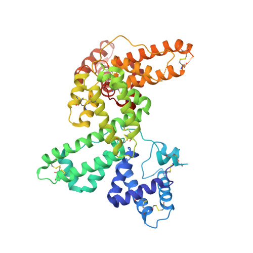
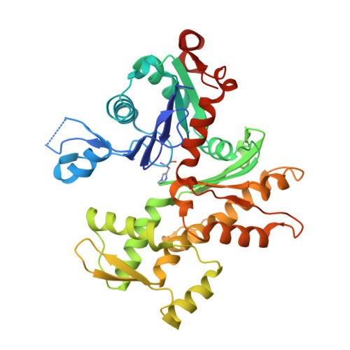
Actin-DBP: the perfect structural fit?

Verboven, C.Bogaerts, I.Waelkens, E.Rabijns, A.Van Baelen, H.Bouillon, R.De Ranter, C.

(2003) Acta Crystallogr D Biol Crystallogr 59: 263-273

  • DOI: https://doi.org/10.1107/s0907444902021455
  • Primary Citation of Related Structures:  
    1MA9

  • PubMed Abstract: 

    The multifunctional vitamin D binding protein (DBP) is an actin-sequestering protein present in blood. The crystal structure of the actin-DBP complex was determined at 2.4 A resolution. DBP binds to actin subdomains 1 and 3 and occludes the cleft at the interface between these subdomains. Most remarkably, DBP demonstrates an unusually large actin-binding interface, far exceeding the binding-interface areas reported for other actin-binding proteins such as profilin, DNase I and gelsolin. The fast-growing side of actin monomers is blocked completely through a perfect structural fit with DBP, demonstrating how DBP effectively interferes with actin-filament formation. It establishes DBP as the hitherto best actin-sequestering protein and highlights its key role in suppressing and preventing extracellular actin polymerization.


  • Organizational Affiliation

    Laboratorium voor Analytische Chemie en Medicinale Fysicochemie, Faculteit Farmaceutische Wetenschappen, K. U. Leuven, Belgium.


Macromolecules
Find similar proteins by:  (by identity cutoff)  |  3D Structure
Entity ID: 1
MoleculeChains Sequence LengthOrganismDetailsImage
Vitamin D-binding protein458Homo sapiensMutation(s): 0 
UniProt & NIH Common Fund Data Resources
Find proteins for P02774 (Homo sapiens)
Explore P02774 
Go to UniProtKB:  P02774
PHAROS:  P02774
GTEx:  ENSG00000145321 
Entity Groups  
Sequence Clusters30% Identity50% Identity70% Identity90% Identity95% Identity100% Identity
UniProt GroupP02774
Sequence Annotations
Expand
  • Reference Sequence
Find similar proteins by:  (by identity cutoff)  |  3D Structure
Entity ID: 2
MoleculeChains Sequence LengthOrganismDetailsImage
Actin, Alpha Skeletal Muscle375Oryctolagus cuniculusMutation(s): 1 
EC: 3.6.4
UniProt
Find proteins for P68135 (Oryctolagus cuniculus)
Explore P68135 
Go to UniProtKB:  P68135
Entity Groups  
Sequence Clusters30% Identity50% Identity70% Identity90% Identity95% Identity100% Identity
UniProt GroupP68135
Sequence Annotations
Expand
  • Reference Sequence
Small Molecules
Modified Residues  1 Unique
IDChains TypeFormula2D DiagramParent
HIC
Query on HIC
B
L-PEPTIDE LINKINGC7 H11 N3 O2HIS
Experimental Data & Validation

Experimental Data

  • Method: X-RAY DIFFRACTION
  • Resolution: 2.40 Å
  • R-Value Free:  0.250 (Depositor), 0.243 (DCC) 
  • R-Value Work:  0.200 (Depositor), 0.194 (DCC) 
  • R-Value Observed: 0.203 (Depositor) 
Space Group: P 1 21 1
Unit Cell:
Length ( Å )Angle ( ˚ )
a = 74.44α = 90
b = 74.9β = 110.19
c = 88.02γ = 90
Software Package:
Software NamePurpose
DENZOdata reduction
SCALEPACKdata scaling
CNSrefinement
CNSphasing

Structure Validation

View Full Validation Report


Ligand Structure Quality Assessment 


Entry History 

Deposition Data

Revision History  (Full details and data files)

  • Version 1.0: 2003-02-04
    Type: Initial release
  • Version 1.1: 2008-04-28
    Changes: Version format compliance
  • Version 1.2: 2011-07-13
    Changes: Version format compliance
  • Version 1.3: 2025-03-26
    Changes: Data collection, Database references, Derived calculations, Refinement description, Structure summary