1M8A

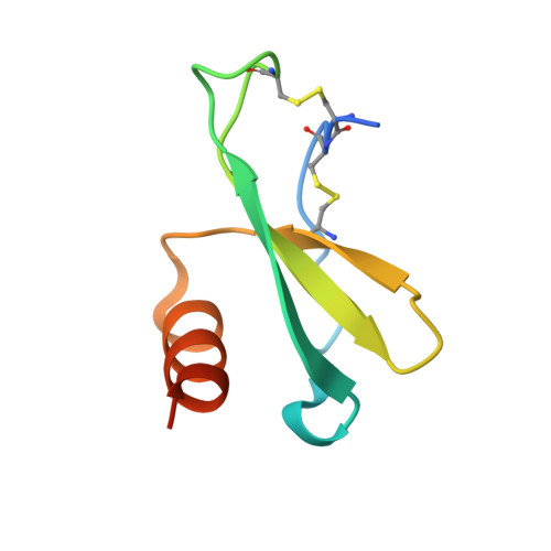
Human MIP-3alpha/CCL20


Experimental Data Snapshot

  • Method: X-RAY DIFFRACTION
  • Resolution: 1.70 Å
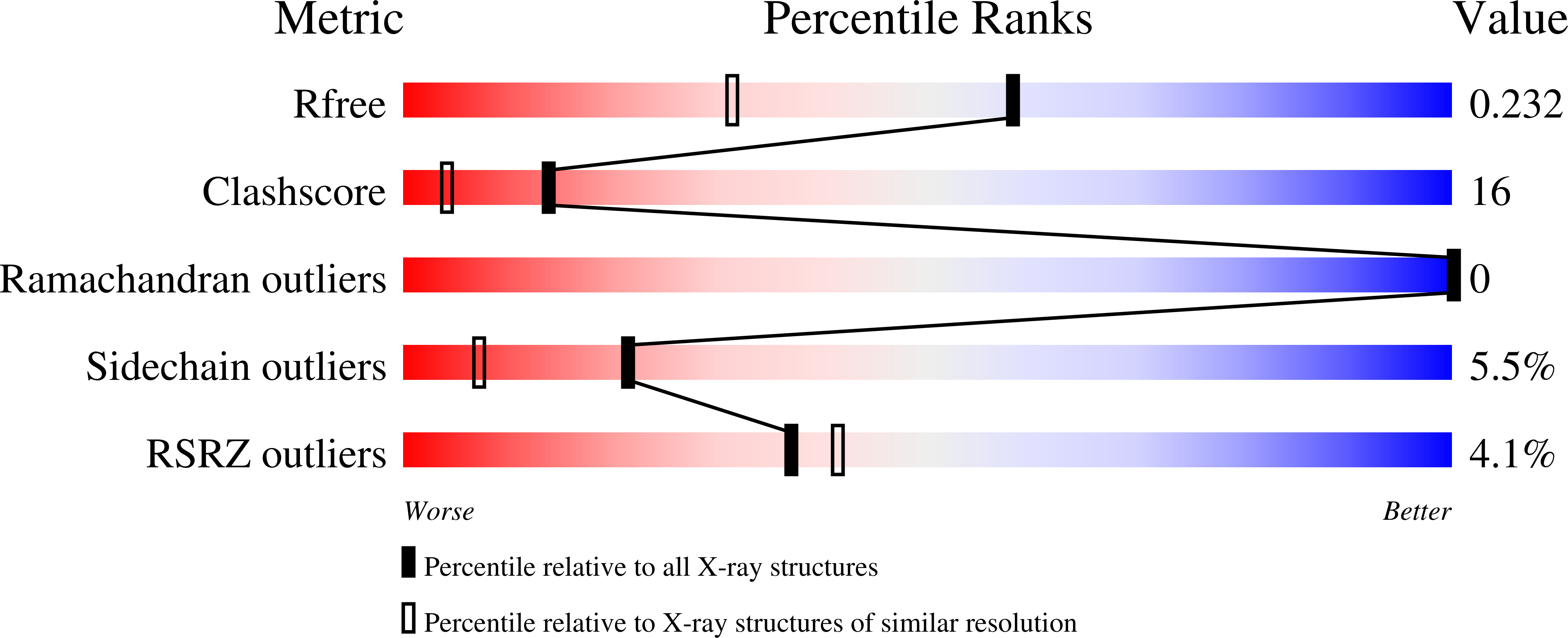
  • R-Value Free: 0.238 
  • R-Value Work: 0.190 
  • R-Value Observed: 0.193 

wwPDB Validation   3D Report Full Report


This is version 1.2 of the entry. See complete history


Literature

The structure of human macrophage inflammatory protein-3alpha /CCL20. Linking antimicrobial and CC chemokine receptor-6-binding activities with human beta-defensins

Hoover, D.M.Boulegue, C.Yang, D.Oppenheim, J.J.Tucker, K.Lu, W.Lubkowski, J.

(2002) J Biol Chem 277: 37647-37654

  • DOI: https://doi.org/10.1074/jbc.M203907200
  • Primary Citation of Related Structures:  
    1M8A

  • PubMed Abstract: 

    Human macrophage inflammatory protein-3alpha (MIP-3alpha; CCL20) is a CC-type chemokine that binds to and activates CC chemokine receptor-6 (CCR6). Although MIP-3alpha does not share the binding site of CCR6 with any other chemokine, human beta-defensin-1 and -2, small cationic antimicrobial peptides, have also been found to bind to and activate CCR6. Conversely, we have found that MIP-3alpha possesses antibacterial activity of greater potency than human beta-defensin-1 and -2 against Escherichia coli ATCC 25922 and Staphylococcus aureus ATCC 29213, while having no activity against the fungus Candida albicans. There is no clear sequence similarity between beta-defensins and the chemokine MIP-3alpha, beyond an abundance of cationic residues and the presence of disulfide bonds. Nonetheless, there are structural similarities between these three proteins that allow their overlap of chemotactic and antimicrobial activities. In this report, we describe the x-ray crystal structure of human MIP-3alpha refined to a resolution of 1.7 A and compare it with the crystal structures of human beta-defensin-1 and -2. Molecules of MIP-3alpha and the beta-defensins seem to share few structural motifs that are likely associated with their common biological activities.


  • Organizational Affiliation

    Macromolecular Crystallography Laboratory, Division of Cancer Treatment, Centers and Diagnosis/Developmental Therapeutics Program, NCI-Frederick, Frederick, Maryland 21702, USA.


Macromolecules
Find similar proteins by:  (by identity cutoff)  |  3D Structure
Entity ID: 1
MoleculeChains Sequence LengthOrganismDetailsImage
Small inducible cytokine A20
A, B
70N/AMutation(s): 0 
UniProt & NIH Common Fund Data Resources
Find proteins for P78556 (Homo sapiens)
Explore P78556 
Go to UniProtKB:  P78556
PHAROS:  P78556
GTEx:  ENSG00000115009 
Entity Groups  
Sequence Clusters30% Identity50% Identity70% Identity90% Identity95% Identity100% Identity
UniProt GroupP78556
Sequence Annotations
Expand
  • Reference Sequence
Small Molecules
Ligands 1 Unique
IDChains Name / Formula / InChI Key2D Diagram3D Interactions
IPA
Query on IPA

Download Ideal Coordinates CCD File 
C [auth A]
D [auth A]
E [auth A]
F [auth A]
G [auth A]
C [auth A],
D [auth A],
E [auth A],
F [auth A],
G [auth A],
H [auth A],
I [auth A],
J [auth B],
K [auth B],
L [auth B]
ISOPROPYL ALCOHOL
C3 H8 O
KFZMGEQAYNKOFK-UHFFFAOYSA-N
Experimental Data & Validation

Experimental Data

  • Method: X-RAY DIFFRACTION
  • Resolution: 1.70 Å
  • R-Value Free: 0.238 
  • R-Value Work: 0.190 
  • R-Value Observed: 0.193 
  • Space Group: P 61
Unit Cell:
Length ( Å )Angle ( ˚ )
a = 70.822α = 90
b = 70.822β = 90
c = 71.768γ = 120
Software Package:
Software NamePurpose
DENZOdata reduction
SCALEPACKdata scaling
AMoREphasing
SHELXL-97refinement

Structure Validation

View Full Validation Report


Entry History 

Deposition Data

Revision History  (Full details and data files)

  • Version 1.0: 2002-07-31
    Type: Initial release
  • Version 1.1: 2008-04-28
    Changes: Version format compliance
  • Version 1.2: 2011-07-13
    Changes: Version format compliance