1M05

HLA B8 in complex with an Epstein Barr Virus determinant


Experimental Data Snapshot

  • Method: X-RAY DIFFRACTION
  • Resolution: 1.90 Å
  • R-Value Free: 0.283 
  • R-Value Work: 0.242 

wwPDB Validation   3D Report Full Report


This is version 1.3 of the entry. See complete history


Literature

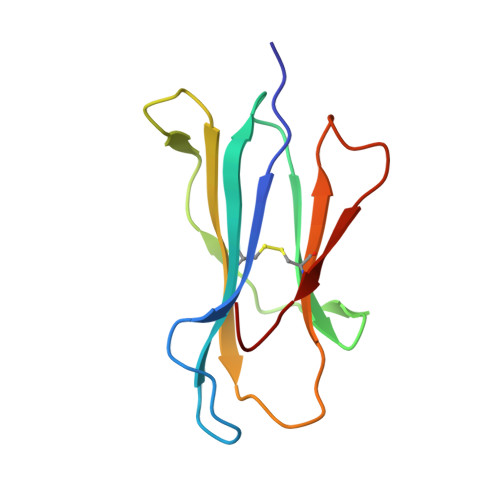
The structure of HLA-B8 complexed to an immunodominant viral determinant: peptide-induced conformational changes and a mode of MHC class I dimerization.

Kjer-Nielsen, L.Clements, C.S.Brooks, A.G.Purcell, A.W.Fontes, M.R.McCluskey, J.Rossjohn, J.

(2003) J Immunol 169: 5153-5160

  • DOI: https://doi.org/10.4049/jimmunol.169.9.5153
  • Primary Citation of Related Structures:  
    1M05

  • PubMed Abstract: 

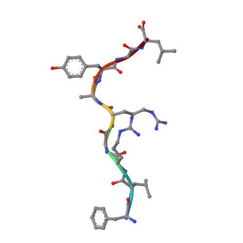
    EBV is a ubiquitous human pathogen that chronically infects up to 90% of the population. Persistent viral infection is characterized by latency and periods of viral replication that are kept in check by a strong antiviral CTL response. Despite the size of the EBV genome, CTL immunity focuses on only a few viral determinants but expands a large primary and memory response toward these epitopes. In unrelated HLA-B8(+) individuals, the response to the immunodominant latent Ag FLRGRAYGL from Epstein Barr nuclear Ag 3A is largely comprised of CTL clones with identical conserved alphabeta TCR structures. To better understand the structural correlates of Ag immunodominance and TCR selection bias, we have solved the crystal structure of the HLA-B8-FLRGRAYGL peptide complex to a resolution of 1.9 A. The structure confirms the importance of P3-Arg, P5-Arg, and P9-Leu as dominant anchor residues involved in peptide binding to HLA-B8. A bulged conformation of the bound peptide provides a structural basis for the critical role of the P7-Tyr residue in T cell recognition. The peptide also induces backbone and side-chain conformational changes in HLA-B8 that are transmitted along the peptide-binding groove in a domino effect. The HLA-B8-FLRGRAYGL complex crystallizes as a dimer in the asymmetric unit and is oriented such that both peptide ligands are projected in the same plane suggesting a higher order arrangement of MHC-peptide complexes that could be involved in formation of the class I Ag-loading complex or in T cell activation.


  • Organizational Affiliation

    Department of Microbiology and Immunology, University of Melbourne, Parkville, Victoria, Australia.


Macromolecules
Find similar proteins by:  (by identity cutoff)  |  3D Structure
Entity ID: 1
MoleculeChains Sequence LengthOrganismDetailsImage
HLA class I histocompatibility antigen, B-8 B*0801 alpha chain
A, C
277Homo sapiensMutation(s): 0 
UniProt & NIH Common Fund Data Resources
Find proteins for P01889 (Homo sapiens)
Explore P01889 
Go to UniProtKB:  P01889
PHAROS:  P01889
GTEx:  ENSG00000234745 
Entity Groups  
Sequence Clusters30% Identity50% Identity70% Identity90% Identity95% Identity100% Identity
UniProt GroupP01889
Sequence Annotations
Expand
  • Reference Sequence
Find similar proteins by:  (by identity cutoff)  |  3D Structure
Entity ID: 2
MoleculeChains Sequence LengthOrganismDetailsImage
Beta-2-microglobulin
B, D
99Homo sapiensMutation(s): 0 
UniProt & NIH Common Fund Data Resources
Find proteins for P61769 (Homo sapiens)
Explore P61769 
Go to UniProtKB:  P61769
PHAROS:  P61769
GTEx:  ENSG00000166710 
Entity Groups  
Sequence Clusters30% Identity50% Identity70% Identity90% Identity95% Identity100% Identity
UniProt GroupP61769
Sequence Annotations
Expand
  • Reference Sequence

Find similar proteins by:  Sequence   |   3D Structure  

Entity ID: 3
MoleculeChains Sequence LengthOrganismDetailsImage
EBNA-3 nuclear protein
E, F
9N/AMutation(s): 0 
UniProt
Find proteins for P12977 (Epstein-Barr virus (strain B95-8))
Explore P12977 
Go to UniProtKB:  P12977
Entity Groups  
UniProt GroupP12977
Sequence Annotations
Expand
  • Reference Sequence
Small Molecules
Ligands 1 Unique
IDChains Name / Formula / InChI Key2D Diagram3D Interactions
CD
Query on CD

Download Ideal Coordinates CCD File 
G [auth B],
H [auth D]
CADMIUM ION
Cd
WLZRMCYVCSSEQC-UHFFFAOYSA-N
Experimental Data & Validation

Experimental Data

  • Method: X-RAY DIFFRACTION
  • Resolution: 1.90 Å
  • R-Value Free: 0.283 
  • R-Value Work: 0.242 
  • Space Group: P 21 21 21
Unit Cell:
Length ( Å )Angle ( ˚ )
a = 85.4α = 90
b = 90.1β = 90
c = 125.3γ = 90
Software Package:
Software NamePurpose
AMoREphasing
CNSrefinement

Structure Validation

View Full Validation Report


Entry History 

Deposition Data

Revision History  (Full details and data files)

  • Version 1.0: 2003-09-02
    Type: Initial release
  • Version 1.1: 2008-04-28
    Changes: Version format compliance
  • Version 1.2: 2011-07-13
    Changes: Version format compliance
  • Version 1.3: 2017-10-11
    Changes: Refinement description