1LXT | pdb_00001lxt

STRUCTURE OF PHOSPHOTRANSFERASE PHOSPHOGLUCOMUTASE FROM RABBIT


Experimental Data Snapshot

  • Method: X-RAY DIFFRACTION
  • Resolution: 2.70 Å
  • R-Value Free: 
    0.260 (Depositor), 0.279 (DCC) 
  • R-Value Work: 
    0.201 (Depositor), 0.246 (DCC) 
  • R-Value Observed: 
    0.201 (Depositor) 

Starting Model: experimental
View more details

wwPDB Validation 3D Report Full Report

Validation slider image for 1LXT

This is version 1.4 of the entry. See complete history

Literature

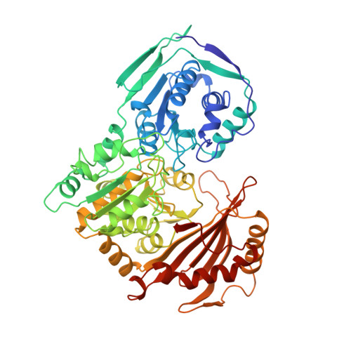
Structure of rabbit muscle phosphoglucomutase refined at 2.4 A resolution.

Liu, Y.Ray, W.J.Baranidharan, S.

(1997) Acta Crystallogr D Biol Crystallogr 53: 392-405

  • DOI: https://doi.org/10.1107/S0907444997000875
  • Primary Citation Related Structures: 
    1LXT, 3PMG

  • PubMed Abstract: 

    Data between 6.0 and 2.4 A resolution, collected at 253 K, wer used to refine a revised atomic model of muscle phosphoglucomutase: final crystallographic R factor = 16.3% (Rfree = 19.1%); final r.m.s. deviations from ideal bond lengths and angles = 0.018 A and 3.2 degrees, respectively. Features of the protein that were recognized only in the revised model include: the disposition of water molecules within domain-domain interfaces; two ion pairs buried in domain-domain interfaces, one of which is a structural arginine around which the active-site phosphoserine loop is wound; the basic architecture of the active-site 'crevice', which is a groove in a 1(1/3)-turn helix, open at both ends, that is produced by the interfacing of the four domains; the distorted hexacoordinate ligand sphere of the active-site Mg2+, where the enzymic phosphate group acts as a bidentate ligand; a pair of arginine residues in domain IV that form part of the enzymic phosphate-binding site (distal subsite) whose disposition in the two monomers of the asymmetric unit is affected unequally by distant crystallographic contacts; structural differences throughout domain IV, produced by these differing contacts, that may mimic solution differences induced by substrate binding; large differences in individually refined Debye-Waller thermal factors for corresponding main-chain atoms in monomers (1) and (2), suggesting a dynamic disorder within the crystal that may involve domain-size groups of residues; and a 'nucleophilic elbow' in the active site that resides in a topological environment differing from previous descriptions of this type of structure in other proteins.


  • Organizational Affiliation
    • The Department of Biological Sciences, Purdue University, West Lafayette, IN 47907, USA.

Macromolecule Content 

  • Total Structure Weight: 123.42 kDa 
  • Atom Count: 9,060 
  • Modeled Residue Count: 1,122 
  • Deposited Residue Count: 1,122 
  • Unique protein chains: 1

Macromolecules

Find similar proteins by:|  3D Structure
Entity ID: 1
MoleculeChains  Sequence LengthOrganismDetailsImage
PHOSPHOGLUCOMUTASE (DEPHOSPHO FORM)
A, B
561Oryctolagus cuniculusMutation(s): 0 
EC: 5.4.2.2
UniProt
Find proteins for P00949 (Oryctolagus cuniculus)
Explore P00949 
Go to UniProtKB:  P00949
Entity Groups
Sequence Clusters30% Identity50% Identity70% Identity90% Identity95% Identity100% Identity
UniProt GroupP00949
Sequence Annotations
Expand
Reference Sequence

Experimental Data & Validation

Experimental Data

  • Method: X-RAY DIFFRACTION
  • Resolution: 2.70 Å
  • R-Value Free:  0.260 (Depositor), 0.279 (DCC) 
  • R-Value Work:  0.201 (Depositor), 0.246 (DCC) 
  • R-Value Observed: 0.201 (Depositor) 
Space Group: P 41 21 2
Unit Cell:
Length ( Å )Angle ( ˚ )
a = 174.42α = 90
b = 174.42β = 90
c = 101.12γ = 90
Software Package:
Software NamePurpose
X-PLORmodel building
X-PLORrefinement
XDSdata reduction
X-PLORphasing

Structure Validation

View Full Validation Report


Entry History 

Deposition Data

Revision History  (Full details and data files)

  • Version 1.0: 1997-02-12
    Type: Initial release
  • Version 1.1: 2008-03-24
    Changes: Version format compliance
  • Version 1.2: 2011-07-13
    Changes: Version format compliance
  • Version 1.3: 2023-08-09
    Changes: Database references, Derived calculations, Other, Refinement description
  • Version 1.4: 2024-05-22
    Changes: Data collection