1LRT

CRYSTAL STRUCTURE OF TERNARY COMPLEX OF TRITRICHOMONAS FOETUS INOSINE-5'-MONOPHOSPHATE DEHYDROGENASE: STRUCTURAL CHARACTERIZATION OF NAD+ SITE IN MICROBIAL ENZYME


Experimental Data Snapshot

  • Method: X-RAY DIFFRACTION
  • Resolution: 2.20 Å
  • R-Value Free: 0.246 
  • R-Value Work: 0.212 
  • R-Value Observed: 0.218 

wwPDB Validation   3D Report Full Report


This is version 1.4 of the entry. See complete history


Literature

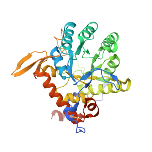
Crystal structure of a ternary complex of Tritrichomonas foetus inosine 5'-monophosphate dehydrogenase: NAD+ orients the active site loop for catalysis

Gan, L.Petsko, G.A.Hedstrom, L.

(2003) Biochemistry 41: 13309-13317

  • DOI: https://doi.org/10.1021/bi0203785
  • Primary Citation of Related Structures:  
    1LRT

  • PubMed Abstract: 

    Inosine 5'-monophosphate dehydrogenase (IMPDH) catalyzes the conversion of IMP to XMP with the reduction of NAD(+), which is the rate-limiting step in the biosynthesis of guanine nucleotides. IMPDH is a promising target for chemotherapy. Microbial IMPDHs differ from mammalian enzymes in their lower affinity for inhibitors such as mycophenolic acid (MPA) and thiazole-4-carboxamide adenine dinucleotide (TAD). Part of this resistance is determined by the coupling between nicotinamide and adenosine subsites in the NAD(+) binding site that is postulated to involve an active site flap. To understand the structural basis of the drug selectivity, we solved the X-ray crystal structure of the catalytic core domain of Tritrichomonas foetus IMPDH in complex with IMP and beta-methylene-TAD at 2.2 A resolution. Unlike previous structures of this enzyme, the active site loop is ordered in this complex, and the catalytic Cys319 is 3.6 A from IMP, in the same plane as the hypoxanthine ring. The active site loop forms hydrogen bonds to the carboxamide of beta-Me-TAD which suggests that NAD(+) promotes the nucleophillic attack of Cys319 on IMP. The interactions of the adenosine end of TAD are very different from those in the human enzyme, suggesting the NAD(+) site may be an exploitable target for the design of antimicrobial drugs. In addition, a new K(+) site is observed at the subunit interface. This site is adjacent to beta-Me-TAD, consistent with the link between the K(+) activation and NAD(+). However, contrary to the coupling model, the flap does not cover the adenosine subsite and remains largely disordered.


  • Organizational Affiliation

    Department of Biochemistry, Brandeis University, Waltham, Massachusetts 02454, USA.


Macromolecules
Find similar proteins by:  (by identity cutoff)  |  3D Structure
Entity ID: 1
MoleculeChains Sequence LengthOrganismDetailsImage
INOSINE-5'-MONOPHOSPHATE DEHYDROGENASE
A, B, C, D
376Tritrichomonas suisMutation(s): 0 
Gene Names: IMPDH
EC: 1.1.1.205
UniProt
Find proteins for P50097 (Tritrichomonas foetus)
Explore P50097 
Go to UniProtKB:  P50097
Entity Groups  
Sequence Clusters30% Identity50% Identity70% Identity90% Identity95% Identity100% Identity
UniProt GroupP50097
Sequence Annotations
Expand
  • Reference Sequence
Small Molecules
Ligands 5 Unique
IDChains Name / Formula / InChI Key2D Diagram3D Interactions
TAD
Query on TAD

Download Ideal Coordinates CCD File 
AA [auth D],
I [auth A],
P [auth B],
V [auth C]
BETA-METHYLENE-THIAZOLE-4-CARBOXYAMIDE-ADENINE DINUCLEOTIDE
C20 H27 N7 O13 P2 S
CRWWKLKZKYLFQV-HVIRUEHBSA-N
IMP
Query on IMP

Download Ideal Coordinates CCD File 
H [auth A],
O [auth B],
U [auth C],
Z [auth D]
INOSINIC ACID
C10 H13 N4 O8 P
GRSZFWQUAKGDAV-KQYNXXCUSA-N
BOG
Query on BOG

Download Ideal Coordinates CCD File 
E [auth A],
F [auth A]
octyl beta-D-glucopyranoside
C14 H28 O6
HEGSGKPQLMEBJL-RKQHYHRCSA-N
TRS
Query on TRS

Download Ideal Coordinates CCD File 
BA [auth D]
CA [auth D]
DA [auth D]
J [auth A]
K [auth A]
BA [auth D],
CA [auth D],
DA [auth D],
J [auth A],
K [auth A],
L [auth A],
M [auth A],
Q [auth B],
R [auth B],
S [auth B],
W [auth C],
X [auth C]
2-AMINO-2-HYDROXYMETHYL-PROPANE-1,3-DIOL
C4 H12 N O3
LENZDBCJOHFCAS-UHFFFAOYSA-O
K
Query on K

Download Ideal Coordinates CCD File 
G [auth A],
N [auth B],
T [auth C],
Y [auth D]
POTASSIUM ION
K
NPYPAHLBTDXSSS-UHFFFAOYSA-N
Binding Affinity Annotations 
IDSourceBinding Affinity
TAD Binding MOAD:  1LRT Ki: 2300 (nM) from 1 assay(s)
PDBBind:  1LRT Ki: 2300 (nM) from 1 assay(s)
Experimental Data & Validation

Experimental Data

  • Method: X-RAY DIFFRACTION
  • Resolution: 2.20 Å
  • R-Value Free: 0.246 
  • R-Value Work: 0.212 
  • R-Value Observed: 0.218 
  • Space Group: P 21 21 21
Unit Cell:
Length ( Å )Angle ( ˚ )
a = 96.144α = 90
b = 112.369β = 90
c = 161.997γ = 90
Software Package:
Software NamePurpose
DENZOdata reduction
SCALEPACKdata scaling
AMoREphasing
CNSrefinement

Structure Validation

View Full Validation Report


Entry History 

Deposition Data

Revision History  (Full details and data files)

  • Version 1.0: 2003-07-07
    Type: Initial release
  • Version 1.1: 2008-04-28
    Changes: Version format compliance
  • Version 1.2: 2011-07-13
    Changes: Version format compliance
  • Version 1.3: 2017-07-26
    Changes: Refinement description, Source and taxonomy
  • Version 1.4: 2020-07-29
    Type: Remediation
    Reason: Carbohydrate remediation
    Changes: Data collection, Derived calculations, Structure summary