1LPD | pdb_00001lpd

HIGH RESOLUTION STRUCTURE OF RECOMBINANT DIANTHIN ANTIVIRAL PROTEIN-POTENT ANTI-HIV AGENT (COMPLEX WITH ADENINE)


Experimental Data Snapshot

  • Method: X-RAY DIFFRACTION
  • Resolution: 1.70 Å
  • R-Value Free: 
    0.230 (Depositor) 
  • R-Value Work: 
    0.170 (Depositor) 
  • R-Value Observed: 
    0.179 (Depositor) 

Starting Model: experimental
View more details

wwPDB Validation 3D Report Full Report

Validation slider image for 1LPD

This is version 1.3 of the entry. See complete history

Literature

High resolution X-ray structure and potent anti-HIV activity of recombinant dianthin antiviral protein.

Kurinov, I.V.Rajamohan, F.Uckun, F.M.

(2004) Arzneimittelforschung 54: 692-702

  • DOI: https://doi.org/10.1055/s-0031-1297024
  • Primary Citation Related Structures: 
    1LP8, 1LPC, 1LPD

  • PubMed Abstract: 

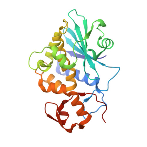
    Dianthin antiviral protein (DAP) is a naturally occurring antiviral protein from the leaves of carnation (Dianthus caryophyllus) capable of depurinating HIV-1 RNA and inhibiting HIV-1 replication in human peripheral blood mononuclear cells. Escherichia coli-derived recombinant DAP (rDAP, amino acids 1-254) was purified to homogeneity for structural and functional studies. In the following paper the X-ray crystal structure of rDAP as well as its complexes with cyclic AMP and adenyl-guanosine (ApG) as substrate analogs at 1.7 A resolution are reported. Molecular modeling studies of the interactions of DAP and the structurally similar pokeweed antiviral protein (PAP) with a single-stranded RNA heptamer predicted a more potent anti-HIV activity for rDAP due to its unique surface topology and more favorable charge distribution in its 20 A-long RNA binding active center cleft. In accordance with the predictions of the modeling studies, rDAP was more potent than rPAP in depurinating HIV-1 RNA. To the knowledge of the authors, this is the first structural and functional characterization of recombinant DAP.


  • Organizational Affiliation
    • Biotherapy and Drug Discovery Program, Parker Hughes Institute, St Paul, Minnesota 55113, USA.

Macromolecule Content 

  • Total Structure Weight: 28.74 kDa 
  • Atom Count: 2,212 
  • Modeled Residue Count: 254 
  • Deposited Residue Count: 254 
  • Unique protein chains: 1

Macromolecules

Find similar proteins by:|  3D Structure
Entity ID: 1
MoleculeChains  Sequence LengthOrganismDetailsImage
Dianthin 30254Dianthus caryophyllusMutation(s): 0 
EC: 3.2.2.22
UniProt
Find proteins for P24476 (Dianthus caryophyllus)
Explore P24476 
Go to UniProtKB:  P24476
Entity Groups
Sequence Clusters30% Identity50% Identity70% Identity90% Identity95% Identity100% Identity
UniProt GroupP24476
Sequence Annotations
Expand
Reference Sequence

Small Molecules

Ligands 1 Unique
IDChains Name / Formula / InChI Key2D Diagram3D Interactions
ADE

Query on ADE


Download:Ideal Coordinates CCD File
B [auth A]ADENINE
C5 H5 N5
GFFGJBXGBJISGV-UHFFFAOYSA-N

Experimental Data & Validation

Experimental Data

  • Method: X-RAY DIFFRACTION
  • Resolution: 1.70 Å
  • R-Value Free:  0.230 (Depositor) 
  • R-Value Work:  0.170 (Depositor) 
  • R-Value Observed: 0.179 (Depositor) 
Space Group: P 1 21 1
Unit Cell:
Length ( Å )Angle ( ˚ )
a = 37.093α = 90
b = 78.464β = 105.26
c = 42.721γ = 90
Software Package:
Software NamePurpose
DENZOdata reduction
SCALEPACKdata scaling
X-PLORmodel building
X-PLORrefinement
X-PLORphasing

Structure Validation

View Full Validation Report


Entry History 

Deposition Data

Revision History  (Full details and data files)

  • Version 1.0: 2004-05-11
    Type: Initial release
  • Version 1.1: 2008-04-28
    Changes: Version format compliance
  • Version 1.2: 2011-07-13
    Changes: Version format compliance
  • Version 1.3: 2023-08-16
    Changes: Data collection, Database references, Derived calculations, Refinement description