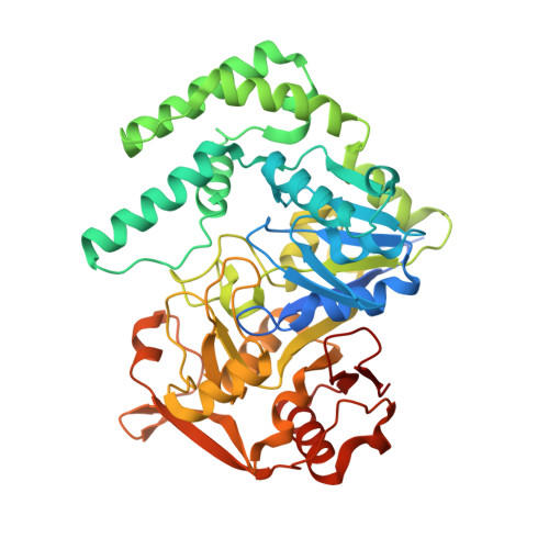
IMP, GTP, and 6-phosphoryl-IMP complexes of recombinant mouse muscle adenylosuccinate synthetase.
Iancu, C.V., Borza, T., Fromm, H.J., Honzatko, R.B.(2002) J Biological Chem 277: 26779-26787
- PubMed: 12004071
- DOI: https://doi.org/10.1074/jbc.M203730200
- Primary Citation of Related Structures:
1IWE, 1LNY, 1LON, 1LOO - PubMed Abstract:
Prokaryotes have a single form of adenylosuccinate synthetase that controls the committed step of AMP biosynthesis, but vertebrates have two isozymes of the synthetase. The basic isozyme, which predominates in muscle, participates in the purine nucleotide cycle, has an active site conformation different from that of the Escherichia coli enzyme, and exhibits significant differences in ligand recognition. Crystalline complexes presented here of the recombinant basic isozyme from mouse show the following. GTP alone binds to the active site without inducing a conformational change. IMP in combination with an acetate anion induces major conformational changes and organizes the active site for catalysis. IMP, in the absence of GTP, binds to the GTP pocket of the synthetase. The combination of GTP and IMP results in the formation of a stable complex of 6-phosphoryl-IMP and GDP in the presence or absence of hadacidin. The response of the basic isozyme to GTP alone differs from that of synthetases from plants, and yet the conformation of the mouse basic and E. coli synthetases in their complexes with GDP, 6-phosphoryl-IMP, and hadacidin are nearly identical. Hence, reported differences in ligand recognition among synthetases probably arise from conformational variations observed in partially ligated enzymes.
- Department of Biochemistry, Biophysics, and Molecular Biology, Molecular Biology Building, Iowa State University, Ames, IA 50011, USA.
Organizational Affiliation:


