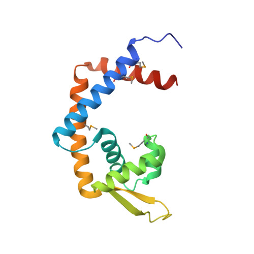
1LNW

CRYSTAL STRUCTURE OF THE MEXR REPRESSOR OF THE MEXAB-OPRM MULTIDRUG EFFLUX OPERON OF PSEUDOMONAS AERUGINOSA


Experimental Data Snapshot

  • Method: X-RAY DIFFRACTION
  • Resolution: 2.10 Å
  • R-Value Free: 0.294 
  • R-Value Work: 0.242 
  • R-Value Observed: 0.242 

wwPDB Validation   3D Report Full Report


This is version 1.3 of the entry. See complete history


Literature

Crystal structure of the MexR repressor of the mexRAB-oprM multidrug efflux operon of Pseudomonas aeruginosa.

Lim, D.Poole, K.Strynadka, N.C.

(2002) J Biol Chem 277: 29253-29259

  • DOI: https://doi.org/10.1074/jbc.M111381200
  • Primary Citation of Related Structures:  
    1LNW

  • PubMed Abstract: 

    MexR is a member of the MarR family of bacterial transcriptional regulators and is the repressor for the MexAB-OprM operon, which encodes a tripartite multidrug efflux system in Pseudomonas aeruginosa. Mutations in MexR result in increased resistance to multiple antibiotics due to overexpression of this efflux system. We have determined the crystal structure of MexR to 2.1-A resolution in the absence of effector. The four copies of the MexR dimer in the asymmetric unit are observed in multiple conformations. Analysis of these conformational states in the context of a model of the MexR-DNA complex proposed in this study suggests that an effector-induced conformational change may inhibit DNA binding by reducing the spacing of the DNA binding domains. The inhibited conformation is exhibited by one of the four MexR dimers, which contains an ordered C-terminal tail from a neighboring monomer inserted between its DNA binding domains and which we propose may resemble the MexR-effector complex. Our results indicate that MexR may differ from the other described member of this family, MarR, in the nature of its effector, mode of DNA binding, and mechanism of regulation.


  • Organizational Affiliation

    Department of Biochemistry and Molecular Biology, University of British Columbia, Vancouver, British Columbia V6T 1Z3, Canada.


Macromolecules
Find similar proteins by:  (by identity cutoff)  |  3D Structure
Entity ID: 1
MoleculeChains Sequence LengthOrganismDetailsImage
Multidrug resistance operon repressor
A, B, C, D, E
A, B, C, D, E, F, G, H
147Pseudomonas aeruginosaMutation(s): 5 
UniProt
Find proteins for P52003 (Pseudomonas aeruginosa (strain ATCC 15692 / DSM 22644 / CIP 104116 / JCM 14847 / LMG 12228 / 1C / PRS 101 / PAO1))
Explore P52003 
Go to UniProtKB:  P52003
Entity Groups  
Sequence Clusters30% Identity50% Identity70% Identity90% Identity95% Identity100% Identity
UniProt GroupP52003
Sequence Annotations
Expand
  • Reference Sequence
Small Molecules
Modified Residues  1 Unique
IDChains TypeFormula2D DiagramParent
MSE
Query on MSE
A, B, C, D, E
A, B, C, D, E, F, G, H
L-PEPTIDE LINKINGC5 H11 N O2 SeMET
Experimental Data & Validation

Experimental Data

  • Method: X-RAY DIFFRACTION
  • Resolution: 2.10 Å
  • R-Value Free: 0.294 
  • R-Value Work: 0.242 
  • R-Value Observed: 0.242 
  • Space Group: P 21 21 21
Unit Cell:
Length ( Å )Angle ( ˚ )
a = 67.91α = 90
b = 72.654β = 90
c = 240.528γ = 90
Software Package:
Software NamePurpose
CNSrefinement
DENZOdata reduction
SCALEPACKdata scaling
CNSphasing

Structure Validation

View Full Validation Report


Entry History 

Deposition Data

Revision History  (Full details and data files)

  • Version 1.0: 2002-09-11
    Type: Initial release
  • Version 1.1: 2008-04-28
    Changes: Version format compliance
  • Version 1.2: 2011-07-13
    Changes: Version format compliance
  • Version 1.3: 2024-10-16
    Changes: Data collection, Database references, Derived calculations, Structure summary