1LIA

CRYSTAL STRUCTURE OF R-PHYCOERYTHRIN FROM POLYSIPHONIA AT 2.8 A RESOLUTION


Experimental Data Snapshot

  • Method: X-RAY DIFFRACTION
  • Resolution: 2.80 Å
  • R-Value Free: 0.182 
  • R-Value Work: 0.180 
  • R-Value Observed: 0.180 

wwPDB Validation   3D Report Full Report


This is version 1.2 of the entry. See complete history


Literature

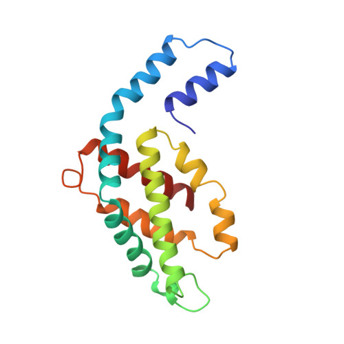
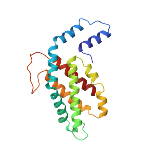
Crystal structure of R-phycoerythrin from Polysiphonia urceolata at 2.8 A resolution.

Chang, W.R.Jiang, T.Wan, Z.L.Zhang, J.P.Yang, Z.X.Liang, D.C.

(1996) J Mol Biol 262: 721-731

  • DOI: https://doi.org/10.1006/jmbi.1996.0547
  • Primary Citation of Related Structures:  
    1LIA

  • PubMed Abstract: 

    The structure of R-phycoerythrin (R-PE) from Polysiphonia urceolata was determined at 2.8 A resolution. The crystals belong to space group R3 with unit cell dimensions of a = b = 189.8 A, c = 60.1 A. The subunit composition of R-PE is (alpha 2 beta 2)3 gamma. The three-dimensional structure of R-PE was solved by the multiple isomorphous replacement method. After several cycles of model building and refinement, the crystallographic R-factor of the final model is 18.0% with data from 10.0 to 2.8 A resolution. The four phycoerythrobilin chromophores alpha 84, alpha 140a, beta 84 and beta 155 in an (alpha beta) unit are each covalently bound to a cysteine residue through ring A. The phycourobilin chromophore is bound to cysteine beta 50 by ring A and bound to cysteine beta 61 by ring D. The ring A and ring D of phycourobilin deviate from the conjugate plane formed by ring B and ring C and the four rings form a boat-shaped structure. R-PE contains a 34 kDa gamma subunit that is assumed to lie in the central channel of the molecular disc (alpha 2 beta 2)3. The energy transfer and relationship between cysteine residues and chromophores are discussed.


  • Organizational Affiliation

    National Laboratory of Biomacromolecules, Chinese Academy of Sciences, Beijing, China.


Macromolecules
Find similar proteins by:  (by identity cutoff)  |  3D Structure
Entity ID: 1
MoleculeChains Sequence LengthOrganismDetailsImage
R-PHYCOERYTHRINA,
C [auth K]
164Polysiphonia urceolataMutation(s): 0 
UniProt
Find proteins for P84861 (Polysiphonia urceolata)
Explore P84861 
Go to UniProtKB:  P84861
Entity Groups  
Sequence Clusters30% Identity50% Identity70% Identity90% Identity95% Identity100% Identity
UniProt GroupP84861
Sequence Annotations
Expand
  • Reference Sequence
Find similar proteins by:  (by identity cutoff)  |  3D Structure
Entity ID: 2
MoleculeChains Sequence LengthOrganismDetailsImage
R-PHYCOERYTHRINB,
D [auth L]
177Polysiphonia urceolataMutation(s): 0 
UniProt
Find proteins for P84862 (Polysiphonia urceolata)
Explore P84862 
Go to UniProtKB:  P84862
Entity Groups  
Sequence Clusters30% Identity50% Identity70% Identity90% Identity95% Identity100% Identity
UniProt GroupP84862
Sequence Annotations
Expand
  • Reference Sequence
Experimental Data & Validation

Experimental Data

  • Method: X-RAY DIFFRACTION
  • Resolution: 2.80 Å
  • R-Value Free: 0.182 
  • R-Value Work: 0.180 
  • R-Value Observed: 0.180 
  • Space Group: H 3
Unit Cell:
Length ( Å )Angle ( ˚ )
a = 189.8α = 90
b = 189.8β = 90
c = 60.1γ = 120
Software Package:
Software NamePurpose
XENGENdata collection
X-PLORmodel building
X-PLORrefinement
XENGENdata reduction
X-PLORphasing

Structure Validation

View Full Validation Report


Entry History 

Deposition Data

Revision History  (Full details and data files)

  • Version 1.0: 1997-07-29
    Type: Initial release
  • Version 1.1: 2008-03-03
    Changes: Version format compliance
  • Version 1.2: 2011-07-13
    Changes: Version format compliance