1LGQ | pdb_00001lgq

Crystal structure of the FHA domain of the Chfr mitotic checkpoint protein


Experimental Data Snapshot

  • Method: X-RAY DIFFRACTION
  • Resolution: 2.10 Å
  • R-Value Free: 
    0.288 (Depositor), 0.289 (DCC) 
  • R-Value Work: 
    0.242 (Depositor), 0.242 (DCC) 

wwPDB Validation 3D Report Full Report

Validation slider image for 1LGQ

This is version 1.4 of the entry. See complete history

Literature

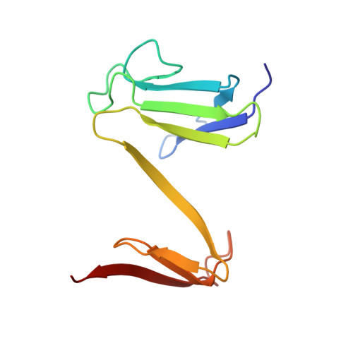
Crystal structure of the FHA domain of the Chfr mitotic checkpoint protein and its complex with tungstate.

Stavridi, E.S.Huyen, Y.Loreto, I.R.Scolnick, D.M.Halazonetis, T.D.Pavletich, N.P.Jeffrey, P.D.

(2002) Structure 10: 891-899

  • DOI: https://doi.org/10.1016/s0969-2126(02)00776-1
  • Primary Citation Related Structures: 
    1LGP, 1LGQ

  • PubMed Abstract: 

    The Chfr mitotic checkpoint protein is frequently inactivated in human cancer. We determined the three-dimensional structure of its FHA domain in its native form and in complex with tungstate, an analog of phosphate. The structures revealed a beta sandwich fold similar to the previously determined folds of the Rad53 N- and C-terminal FHA domains, except that the Rad53 domains were monomeric, whereas the Chfr FHA domain crystallized as a segment-swapped dimer. The ability of the Chfr FHA domain to recognize tungstate suggests that it shares the ability with other FHA domains to bind phosphoproteins. Nevertheless, differences in the sequence and structure of the Chfr and Rad53 FHA domains suggest that FHA domains can be divided into families with distinct binding properties.


  • Organizational Affiliation
    • Molecular Genetics Program, Structural Biology Program, Wistar Institute, Philadelphia, PA 19104, USA.

Macromolecule Content 

  • Total Structure Weight: 25.52 kDa 
  • Atom Count: 1,934 
  • Modeled Residue Count: 224 
  • Deposited Residue Count: 224 
  • Unique protein chains: 1

Macromolecules

Find similar proteins by:|  3D Structure
Entity ID: 1
MoleculeChains  Sequence LengthOrganismDetailsImage
cell cycle checkpoint protein CHFR
A, B
112Homo sapiensMutation(s): 0 
EC: 2.3.2.27
UniProt & NIH Common Fund Data Resources
Find proteins for Q96EP1 (Homo sapiens)
Explore Q96EP1 
Go to UniProtKB:  Q96EP1
PHAROS:  Q96EP1
GTEx:  ENSG00000072609 
Entity Groups
Sequence Clusters30% Identity50% Identity70% Identity90% Identity95% Identity100% Identity
UniProt GroupQ96EP1
Sequence Annotations
Expand
Reference Sequence

Experimental Data & Validation

Experimental Data

  • Method: X-RAY DIFFRACTION
  • Resolution: 2.10 Å
  • R-Value Free:  0.288 (Depositor), 0.289 (DCC) 
  • R-Value Work:  0.242 (Depositor), 0.242 (DCC) 
Space Group: P 21 21 21
Unit Cell:
Length ( Å )Angle ( ˚ )
a = 54.9α = 90
b = 52.9β = 90
c = 77.25γ = 90
Software Package:
Software NamePurpose
CNSrefinement
HKL-2000data reduction
SCALEPACKdata scaling
CNSphasing

Structure Validation

View Full Validation Report


Entry History 

Deposition Data

Revision History  (Full details and data files)

  • Version 1.0: 2002-05-08
    Type: Initial release
  • Version 1.1: 2008-04-28
    Changes: Version format compliance
  • Version 1.2: 2011-07-13
    Changes: Version format compliance
  • Version 1.3: 2022-12-21
    Changes: Database references
  • Version 1.4: 2024-05-22
    Changes: Data collection