1LF6

CRYSTAL STRUCTURE OF BACTERIAL GLUCOAMYLASE


Experimental Data Snapshot

  • Method: X-RAY DIFFRACTION
  • Resolution: 2.10 Å
  • R-Value Free: 0.232 
  • R-Value Work: 0.194 

wwPDB Validation   3D Report Full Report


This is version 1.4 of the entry. See complete history


Literature

Crystal structure and evolution of prokaryotic glucoamylase

Aleshin, A.E.Feng, P.-H.Honzatko, R.B.Reilly, P.J.

(2003) J Mol Biol 327: 61-73

  • DOI: https://doi.org/10.1016/s0022-2836(03)00084-6
  • Primary Citation of Related Structures:  
    1LF6, 1LF9

  • PubMed Abstract: 

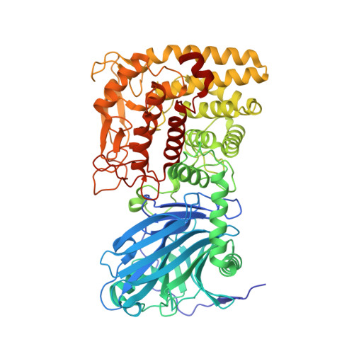
    The first crystal structures of a two-domain, prokaryotic glucoamylase were determined to high resolution from the clostridial species Thermoanaerobacterium thermosaccharolyticum with and without acarbose. The N-terminal domain has 18 antiparallel strands arranged in beta-sheets of a super-beta-sandwich. The C-terminal domain is an (alpha/alpha)(6) barrel, lacking the peripheral subdomain of eukaryotic glucoamylases. Interdomain contacts are common to all prokaryotic Family GH15 proteins. Domains similar to those of prokaryotic glucoamylases in maltose phosphorylases (Family GH65) and glycoaminoglycan lyases (Family PL8) suggest evolution from a common ancestor. Eukaryotic glucoamylases may have evolved from prokaryotic glucoamylases by the substitution of the N-terminal domain with the peripheral subdomain and by the addition of a starch-binding domain.


  • Organizational Affiliation

    Department of Biochemistry, Biophysics, and Molecular Biology, Iowa State University, Ames, IA 50011, USA.


Macromolecules
Find similar proteins by:  (by identity cutoff)  |  3D Structure
Entity ID: 1
MoleculeChains Sequence LengthOrganismDetailsImage
glucoamylase
A, B
684Thermoanaerobacterium thermosaccharolyticumMutation(s): 0 
EC: 3.2.1.3
UniProt
Find proteins for O85672 (Thermoanaerobacterium thermosaccharolyticum)
Explore O85672 
Go to UniProtKB:  O85672
Entity Groups  
Sequence Clusters30% Identity50% Identity70% Identity90% Identity95% Identity100% Identity
UniProt GroupO85672
Sequence Annotations
Expand
  • Reference Sequence
Experimental Data & Validation

Experimental Data

  • Method: X-RAY DIFFRACTION
  • Resolution: 2.10 Å
  • R-Value Free: 0.232 
  • R-Value Work: 0.194 
  • Space Group: P 21 21 21
Unit Cell:
Length ( Å )Angle ( ˚ )
a = 81.186α = 90
b = 102.513β = 90
c = 164.82γ = 90
Software Package:
Software NamePurpose
DENZOdata reduction
SCALEPACKdata scaling
SOLVEphasing
CNSrefinement

Structure Validation

View Full Validation Report


Entry History 

Deposition Data

Revision History  (Full details and data files)

  • Version 1.0: 2003-02-25
    Type: Initial release
  • Version 1.1: 2008-04-28
    Changes: Version format compliance
  • Version 1.2: 2011-07-13
    Changes: Version format compliance
  • Version 1.3: 2018-01-31
    Changes: Experimental preparation
  • Version 1.4: 2024-02-14
    Changes: Data collection, Database references, Derived calculations