1LEN

REFINEMENT OF TWO CRYSTAL FORMS OF LENTIL LECTIN AT 1.8 ANGSTROMS RESOLUTION


Experimental Data Snapshot

  • Method: X-RAY DIFFRACTION
  • Resolution: 1.80 Å
  • R-Value Observed: 0.175 

wwPDB Validation   3D Report Full Report


This is version 1.5 of the entry. See complete history


Literature

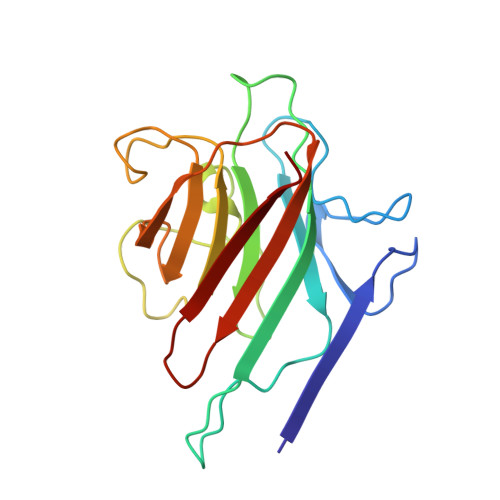
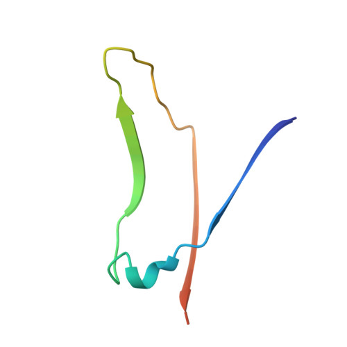
Structural analysis of two crystal forms of lentil lectin at 1.8 A resolution.

Loris, R.Van Overberge, D.Dao-Thi, M.H.Poortmans, F.Maene, N.Wyns, L.

(1994) Proteins 20: 330-346

  • DOI: https://doi.org/10.1002/prot.340200406
  • Primary Citation of Related Structures:  
    1LEN, 2LAL

  • PubMed Abstract: 

    The structures of two crystal forms of lentil lectin are determined and refined at high resolution. Orthorhombic lentil lectin is refined at 1.80 A resolution to an R-factor of 0.184 and monoclinic lentil lectin at 1.75 A resolution to an R-factor of 0.175. These two structures are compared to each other and to the other available legume lectin structures. The monosaccharide binding pocket of each lectin monomer contains a tightly bound phosphate ion. This phosphate makes hydrogen bonding contacts with Asp-81 beta, Gly-99 beta, and Asn-125 beta, three residues that are highly conserved in most of the known legume lectin sequences and essential for monosaccharide recognition in all legume lectin crystal structures described thus far. A detailed analysis of the composition and properties of the hydrophobic contact network and hydrophobic nuclei in lentil lectin is presented. Contact map calculations reveal that dense clusters of nonpolar as well as polar side chains play a major role in secondary structure packing. This is illustrated by a large cluster of 24 mainly hydrophobic amino acids that is responsible for the majority of packing interactions between the two beta-sheets. Another series of four smaller and less hydrophobic clusters is found to mediate the packing of a number of loop structures upon the front sheet. A very dense, but not very conserved cluster is found to stabilize the transition metal binding site. The highly conserved and invariant nonpolar residues are distributed asymmetrically over the protein.


  • Organizational Affiliation

    Laboratorium voor Ultrastructuur, Vrije Universiteit Brussel, Sint-Genesius-Rode, Belgium.


Macromolecules
Find similar proteins by:  (by identity cutoff)  |  3D Structure
Entity ID: 1
MoleculeChains Sequence LengthOrganismDetailsImage
LECTIN
A, C
181Lens culinarisMutation(s): 0 
UniProt
Find proteins for P02870 (Lens culinaris)
Explore P02870 
Go to UniProtKB:  P02870
Entity Groups  
Sequence Clusters30% Identity50% Identity70% Identity90% Identity95% Identity100% Identity
UniProt GroupP02870
Sequence Annotations
Expand
  • Reference Sequence
Find similar proteins by:  (by identity cutoff)  |  3D Structure
Entity ID: 2
MoleculeChains Sequence LengthOrganismDetailsImage
LECTIN
B, D
52Lens culinarisMutation(s): 0 
UniProt
Find proteins for P02870 (Lens culinaris)
Explore P02870 
Go to UniProtKB:  P02870
Entity Groups  
Sequence Clusters30% Identity50% Identity70% Identity90% Identity95% Identity100% Identity
UniProt GroupP02870
Sequence Annotations
Expand
  • Reference Sequence
Experimental Data & Validation

Experimental Data

  • Method: X-RAY DIFFRACTION
  • Resolution: 1.80 Å
  • R-Value Observed: 0.175 
  • Space Group: P 1 21 1
Unit Cell:
Length ( Å )Angle ( ˚ )
a = 58.49α = 90
b = 56.37β = 104.4
c = 82.73γ = 90
Software Package:
Software NamePurpose
RESTRAINrefinement

Structure Validation

View Full Validation Report


Entry History 

Deposition Data

Revision History  (Full details and data files)

  • Version 1.0: 1994-01-31
    Type: Initial release
  • Version 1.1: 2008-03-03
    Changes: Version format compliance
  • Version 1.2: 2011-07-13
    Changes: Version format compliance
  • Version 1.3: 2017-11-29
    Changes: Derived calculations, Other
  • Version 1.4: 2020-09-09
    Changes: Database references, Derived calculations
  • Version 1.5: 2024-02-14
    Changes: Data collection, Database references