1LAG

STRUCTURAL BASES FOR MULTIPLE LIGAND SPECIFICITY OF THE PERIPLASMIC LYSINE-, ARGININE-, ORNITHINE-BINDING PROTEIN


Experimental Data Snapshot

  • Method: X-RAY DIFFRACTION
  • Resolution: 2.06 Å
  • R-Value Work: 0.162 
  • R-Value Observed: 0.162 

wwPDB Validation   3D Report Full Report


This is version 1.3 of the entry. See complete history


Literature

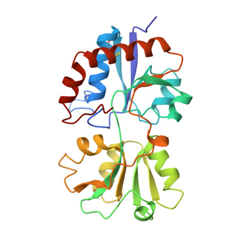
Structural basis for multiple ligand specificity of the periplasmic lysine-, arginine-, ornithine-binding protein.

Oh, B.H.Ames, G.F.Kim, S.H.

(1994) J Biol Chem 269: 26323-26330

  • Primary Citation of Related Structures:  
    1LAF, 1LAG, 1LAH

  • PubMed Abstract: 

    The substrate-binding site of a protein with multiple specificity should satisfy geometric and energetic complementarity for several different substrates. The structural basis of the multiple ligand specificity of the periplasmic lysine-, arginine-, ornithine-binding protein (LAO) was investigated by determining and analyzing the structures of the protein unliganded and liganded with each of the three high-affinity ligands (L-lysine, L-arginine, and L-ornithine) and with one low-affinity ligand (L-histidine). The geometric complementarity is achieved primarily by virtue of the large size of the ligand-binding site which can accommodate the maximum common volume of the four ligands plus three water molecules. The optimization of energetic complementarity is achieved by the relocation of protein-bound water molecules and by the movement of the Asp-11 side chain. The structure of the LAO-histidine complex indicates that the 30-fold reduced affinity of the protein for histidine is primarily due to unavailability of one ionic interaction of the histidine side chain with the protein which is present in the other three complexes.


  • Organizational Affiliation

    Department of Chemistry, Lawrence Berkeley Laboratory, Berkeley, California.


Macromolecules
Find similar proteins by:  (by identity cutoff)  |  3D Structure
Entity ID: 1
MoleculeChains Sequence LengthOrganismDetailsImage
LYSINE, ARGININE, ORNITHINE-BINDING PROTEINA [auth E]238Salmonella enterica subsp. enterica serovar TyphimuriumMutation(s): 0 
UniProt
Find proteins for P02911 (Salmonella typhimurium (strain LT2 / SGSC1412 / ATCC 700720))
Explore P02911 
Go to UniProtKB:  P02911
Entity Groups  
Sequence Clusters30% Identity50% Identity70% Identity90% Identity95% Identity100% Identity
UniProt GroupP02911
Sequence Annotations
Expand
  • Reference Sequence
Small Molecules
Ligands 1 Unique
IDChains Name / Formula / InChI Key2D Diagram3D Interactions
HIS
Query on HIS

Download Ideal Coordinates CCD File 
B [auth E]HISTIDINE
C6 H10 N3 O2
HNDVDQJCIGZPNO-YFKPBYRVSA-O
Binding Affinity Annotations 
IDSourceBinding Affinity
HIS Binding MOAD:  1LAG Kd: 500 (nM) from 1 assay(s)
PDBBind:  1LAG Kd: 500 (nM) from 1 assay(s)
Experimental Data & Validation

Experimental Data

  • Method: X-RAY DIFFRACTION
  • Resolution: 2.06 Å
  • R-Value Work: 0.162 
  • R-Value Observed: 0.162 
  • Space Group: P 21 21 21
Unit Cell:
Length ( Å )Angle ( ˚ )
a = 37.49α = 90
b = 58.19β = 90
c = 117.65γ = 90
Software Package:
Software NamePurpose
X-PLORmodel building
X-PLORrefinement
X-PLORphasing

Structure Validation

View Full Validation Report


Entry History 

Deposition Data

Revision History  (Full details and data files)

  • Version 1.0: 1995-07-10
    Type: Initial release
  • Version 1.1: 2008-03-24
    Changes: Version format compliance
  • Version 1.2: 2011-07-13
    Changes: Version format compliance
  • Version 1.3: 2017-11-29
    Changes: Derived calculations, Other