1L9M

Three-dimensional structure of the human transglutaminase 3 enzyme: binding of calcium ions change structure for activation


Experimental Data Snapshot

  • Method: X-RAY DIFFRACTION
  • Resolution: 2.10 Å
  • R-Value Free: 0.225 
  • R-Value Work: 0.182 
  • R-Value Observed: 0.296 

wwPDB Validation   3D Report Full Report


This is version 1.4 of the entry. See complete history


Literature

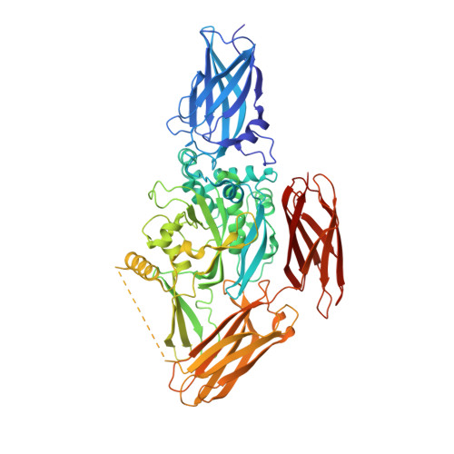
Three-dimensional structure of the human transglutaminase 3 enzyme: binding of calcium ions changes structure for activation.

Ahvazi, B.Kim, H.C.Kee, S.H.Nemes, Z.Steinert, P.M.

(2002) EMBO J 21: 2055-2067

  • DOI: https://doi.org/10.1093/emboj/21.9.2055
  • Primary Citation of Related Structures:  
    1L9M, 1L9N

  • PubMed Abstract: 

    Transglutaminase (TGase) enzymes catalyze the formation of covalent cross-links between protein-bound glutamines and lysines in a calcium-dependent manner, but the role of Ca(2+) ions remains unclear. The TGase 3 isoform is widely expressed and is important for epithelial barrier formation. It is a zymogen, requiring proteolysis for activity. We have solved the three-dimensional structures of the zymogen and the activated forms at 2.2 and 2.1 A resolution, respectively, and examined the role of Ca(2+) ions. The zymogen binds one ion tightly that cannot be exchanged. Upon proteolysis, the enzyme exothermally acquires two more Ca(2+) ions that activate the enzyme, are exchangeable and are functionally replaceable by other lanthanide trivalent cations. Binding of a Ca(2+) ion at one of these sites opens a channel which exposes the key Trp236 and Trp327 residues that control substrate access to the active site. Together, these biochemical and structural data reveal for the first time in a TGase enzyme that Ca(2+) ions induce structural changes which at least in part dictate activity and, moreover, may confer substrate specificity.


  • Organizational Affiliation

    Laboratory of Skin Biology, National Institute of Arthritis and Musculoskeletal and Skin Diseases, National Institutes of Health, Bethesda, MD 20892-8023, USA. Bijan@discus.niams.nih.gov


Macromolecules
Find similar proteins by:  (by identity cutoff)  |  3D Structure
Entity ID: 1
MoleculeChains Sequence LengthOrganismDetailsImage
Protein-glutamine glutamyltransferase E3
A, B
692Homo sapiensMutation(s): 1 
Gene Names: TGM3
EC: 2.3.2.13
UniProt & NIH Common Fund Data Resources
Find proteins for Q08188 (Homo sapiens)
Explore Q08188 
Go to UniProtKB:  Q08188
PHAROS:  Q08188
GTEx:  ENSG00000125780 
Entity Groups  
Sequence Clusters30% Identity50% Identity70% Identity90% Identity95% Identity100% Identity
UniProt GroupQ08188
Sequence Annotations
Expand
  • Reference Sequence
Experimental Data & Validation

Experimental Data

  • Method: X-RAY DIFFRACTION
  • Resolution: 2.10 Å
  • R-Value Free: 0.225 
  • R-Value Work: 0.182 
  • R-Value Observed: 0.296 
  • Space Group: P 1
Unit Cell:
Length ( Å )Angle ( ˚ )
a = 57.709α = 97.24
b = 67.515β = 90.15
c = 116.208γ = 98.67
Software Package:
Software NamePurpose
DENZOdata reduction
SCALEPACKdata scaling
AMoREphasing
CNSrefinement

Structure Validation

View Full Validation Report


Entry History 

Deposition Data

  • Released Date: 2002-05-29 
  • Deposition Author(s): Ahvazi, B.

Revision History  (Full details and data files)

  • Version 1.0: 2002-05-29
    Type: Initial release
  • Version 1.1: 2008-04-28
    Changes: Version format compliance
  • Version 1.2: 2011-07-13
    Changes: Version format compliance
  • Version 1.3: 2021-10-27
    Changes: Advisory, Database references, Derived calculations
  • Version 1.4: 2023-08-16
    Changes: Data collection, Refinement description