Biophysical studies of eIF4E cap-binding protein: recognition of mRNA 5' cap structure and synthetic fragments of eIF4G and 4E-BP1 proteins.
Niedzwiecka, A., Marcotrigiano, J., Stepinski, J., Jankowska-Anyszka, M., Wyslouch-Cieszynska, A., Dadlez, M., Gingras, A.C., Mak, P., Darzynkiewicz, E., Sonenberg, N., Burley, S.K., Stolarski, R.(2002) J Mol Biology 319: 615-635
- PubMed: 12054859 Search on PubMed
- DOI: https://doi.org/10.1016/S0022-2836(02)00328-5
- Primary Citation Related Structures:
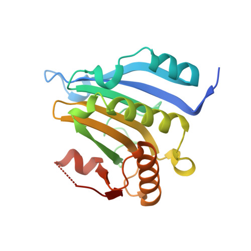
1L8B - PubMed Abstract:
mRNA 5'-cap recognition by the eukaryotic translation initiation factor eIF4E has been exhaustively characterized with the aid of a novel fluorometric, time-synchronized titration method, and X-ray crystallography. The association constant values of recombinant eIF4E for 20 different cap analogues cover six orders of magnitude; with the highest affinity observed for m(7)GTP (approximately 1.1 x 10(8) M(-1)). The affinity of the cap analogues for eIF4E correlates with their ability to inhibit in vitro translation. The association constants yield contributions of non-covalent interactions involving single structural elements of the cap to the free energy of binding, giving a reliable starting point to rational drug design. The free energy of 7-methylguanine stacking and hydrogen bonding (-4.9 kcal/mol) is separate from the energies of phosphate chain interactions (-3.0, -1.9, -0.9 kcal/mol for alpha, beta, gamma phosphates, respectively), supporting two-step mechanism of the binding. The negatively charged phosphate groups of the cap act as a molecular anchor, enabling further formation of the intermolecular contacts within the cap-binding slot. Stabilization of the stacked Trp102/m(7)G/Trp56 configuration is a precondition to form three hydrogen bonds with Glu103 and Trp102. Electrostatically steered eIF4E-cap association is accompanied by additional hydration of the complex by approximately 65 water molecules, and by ionic equilibria shift. Temperature dependence reveals the enthalpy-driven and entropy-opposed character of the m(7)GTP-eIF4E binding, which results from dominant charge-related interactions (DeltaH degrees =-17.8 kcal/mol, DeltaS degrees= -23.6 cal/mol K). For recruitment of synthetic eIF4GI, eIF4GII, and 4E-BP1 peptides to eIF4E, all the association constants were approximately 10(7) M(-1), in decreasing order: eIF4GI>4E-BP1>eIF4GII approximately 4E-BP1(P-Ser65) approximately 4E-BP1(P-Ser65/Thr70). Phosphorylation of 4E-BP1 at Ser65 and Thr70 is insufficient to prevent binding to eIF4E. Enhancement of the eIF4E affinity for cap occurs after binding to eIF4G peptides.
- Department of Biophysics, Institute of Experimental Physics, Warsaw University, 93 Zwirki & Wigury Street, 02-089 Warsaw, Poland.
Organizational Affiliation: