1KP0

The Crystal Structure Analysis of Creatine Amidinohydrolase from Actinobacillus


Experimental Data Snapshot

  • Method: X-RAY DIFFRACTION
  • Resolution: 2.70 Å
  • R-Value Free: 0.222 
  • R-Value Work: 0.188 
  • R-Value Observed: 0.191 

wwPDB Validation   3D Report Full Report


This is version 1.3 of the entry. See complete history


Literature

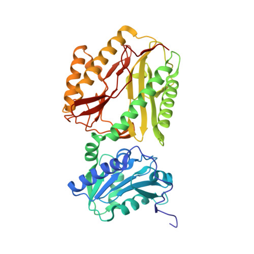
Structure of creatine amidinohydrolase from Actinobacillus.

Padmanabhan, B.Paehler, A.Horikoshi, M.

(2002) Acta Crystallogr D Biol Crystallogr 58: 1322-1328

  • DOI: https://doi.org/10.1107/s0907444902010156
  • Primary Citation of Related Structures:  
    1KP0

  • PubMed Abstract: 

    The crystal structure of Actinobacillus creatine amidinohydrolase has been solved by molecular replacement. The amino-acid sequence has been derived from the crystal structure. Crystals belong to space group I222, with unit-cell parameters a = 111.26 (3), b = 113.62 (4), c = 191.65 (2) A, and contain two molecules in an asymmetric unit. The structure was refined to an R factor of 18.8% at 2.7 A resolution. The crystal structure contains a dimer of 402 residues and 118 water molecules. The protein structure is bilobal, consisting of a small N-terminal domain and a large C-terminal domain. The C-terminal domain has a pitta-bread fold, similar to that found in Pseudomonas putida creatinase, proline aminopeptidases and methionine aminopeptidase. Comparison with complex crystal structures of P. putida creatinase reveals that the enzyme activity of Actinobacillus creatinase might be similar to that of P. putida creatinase.


  • Organizational Affiliation

    Horikoshi Gene Selector Project, Exploratory Research for Advanced Technology (ERATO), Japan Science and Technology Corporation (JST), 5-9-6 Tokodai, Tsukuba, Ibaraki 300-2635, Japan.


Macromolecules
Find similar proteins by:  (by identity cutoff)  |  3D Structure
Entity ID: 1
MoleculeChains Sequence LengthOrganismDetailsImage
CREATINE AMIDINOHYDROLASE
A, B
402ActinobacillusMutation(s): 0 
EC: 3.5.3.3
UniProt
Find proteins for Q7SIB5 (Actinobacillus)
Explore Q7SIB5 
Go to UniProtKB:  Q7SIB5
Entity Groups  
Sequence Clusters30% Identity50% Identity70% Identity90% Identity95% Identity100% Identity
UniProt GroupQ7SIB5
Sequence Annotations
Expand
  • Reference Sequence
Experimental Data & Validation

Experimental Data

  • Method: X-RAY DIFFRACTION
  • Resolution: 2.70 Å
  • R-Value Free: 0.222 
  • R-Value Work: 0.188 
  • R-Value Observed: 0.191 
  • Space Group: I 2 2 2
Unit Cell:
Length ( Å )Angle ( ˚ )
a = 111.263α = 90
b = 113.624β = 90
c = 191.649γ = 90
Software Package:
Software NamePurpose
AMoREphasing
CNSrefinement
DPSdata reduction
MOSFLMdata reduction

Structure Validation

View Full Validation Report


Entry History 

Deposition Data

Revision History  (Full details and data files)

  • Version 1.0: 2002-07-31
    Type: Initial release
  • Version 1.1: 2008-04-27
    Changes: Version format compliance
  • Version 1.2: 2011-07-13
    Changes: Version format compliance
  • Version 1.3: 2023-08-16
    Changes: Data collection, Database references, Refinement description