1KKO | pdb_00001kko

CRYSTAL STRUCTURE OF CITROBACTER AMALONATICUS METHYLASPARTATE AMMONIA LYASE


Experimental Data Snapshot

  • Method: X-RAY DIFFRACTION
  • Resolution: 1.33 Å
  • R-Value Free: 
    0.191 (Depositor) 
  • R-Value Work: 
    0.161 (Depositor) 
  • R-Value Observed: 
    0.162 (Depositor) 

wwPDB Validation 3D Report Full Report

Validation slider image for 1KKO

This is version 1.3 of the entry. See complete history

Literature

Insights into enzyme evolution revealed by the structure of methylaspartate ammonia lyase.

Levy, C.W.Buckley, P.A.Sedelnikova, S.Kato, Y.Asano, Y.Rice, D.W.Baker, P.J.

(2002) Structure 10: 105-113

  • DOI: https://doi.org/10.1016/s0969-2126(01)00696-7
  • Primary Citation Related Structures: 
    1KKO, 1KKR

  • PubMed Abstract: 

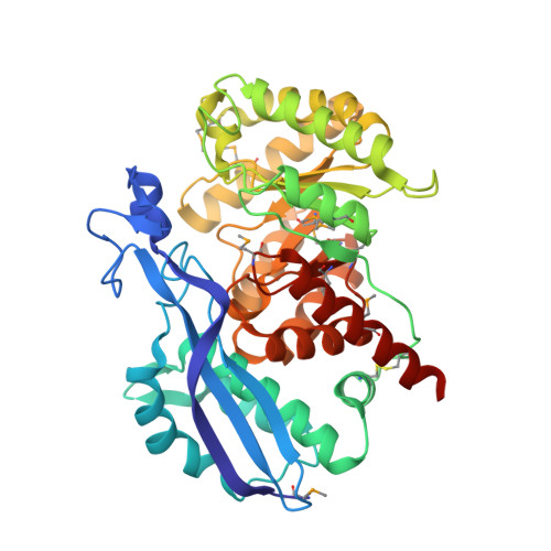
    Methylaspartate ammonia lyase (MAL) catalyzes the magnesium-dependent reversible alpha,beta-elimination of ammonia from L-threo-(2S,3S)-3-methylaspartic acid to mesaconic acid. The 1.3 A MAD crystal structure of the dimeric Citrobacter amalonaticus MAL shows that each subunit comprises two domains, one of which adopts the classical TIM barrel fold, with the active site at the C-terminal end of the barrel. Despite very low sequence similarity, the structure of MAL is closely related to those of representative members of the enolase superfamily, indicating that the mechanism of MAL involves the initial abstraction of a proton alpha to the 3-carboxyl of (2S,3S)-3-methylasparic acid to yield an enolic intermediate. This analysis resolves the conflict that had linked MAL to the histidine and phenylalanine ammonia lyase family of enzymes.


  • Organizational Affiliation
    • Krebs Institute for Biomolecular Research, Department of Molecular Biology and Biotechnology, The University of Sheffield, Sheffield S10 2TN, United Kingdom.

Macromolecule Content 

  • Total Structure Weight: 92.08 kDa 
  • Atom Count: 8,530 
  • Modeled Residue Count: 822 
  • Deposited Residue Count: 826 
  • Unique protein chains: 1

Macromolecules

Find similar proteins by:|  3D Structure
Entity ID: 1
MoleculeChains  Sequence LengthOrganismDetailsImage
3-METHYLASPARTATE AMMONIA-LYASE
A, B
413Citrobacter amalonaticusMutation(s): 10 
Gene Names: MAL
EC: 4.3.1.2
UniProt
Find proteins for O66145 (Citrobacter amalonaticus)
Explore O66145 
Go to UniProtKB:  O66145
Entity Groups
Sequence Clusters30% Identity50% Identity70% Identity90% Identity95% Identity100% Identity
UniProt GroupO66145
Sequence Annotations
Expand
Reference Sequence

Small Molecules

Ligands 1 Unique
IDChains Name / Formula / InChI Key2D Diagram3D Interactions
SO4

Query on SO4


Download:Ideal Coordinates CCD File
C [auth A]SULFATE ION
O4 S
QAOWNCQODCNURD-UHFFFAOYSA-L
Modified Residues  1 Unique
IDChains TypeFormula2D DiagramParent
MSE
Query on MSE
A, B
L-PEPTIDE LINKINGC5 H11 N O2 SeMET

Experimental Data & Validation

Experimental Data

  • Method: X-RAY DIFFRACTION
  • Resolution: 1.33 Å
  • R-Value Free:  0.191 (Depositor) 
  • R-Value Work:  0.161 (Depositor) 
  • R-Value Observed: 0.162 (Depositor) 
Space Group: C 2 2 2
Unit Cell:
Length ( Å )Angle ( ˚ )
a = 128.339α = 90
b = 237.362β = 90
c = 65.806γ = 90
Software Package:
Software NamePurpose
DENZOdata reduction
SCALEPACKdata scaling
CNSrefinement
REFMACrefinement
CNSphasing

Structure Validation

View Full Validation Report


Entry History 

Deposition Data

Revision History  (Full details and data files)

  • Version 1.0: 2002-01-30
    Type: Initial release
  • Version 1.1: 2008-04-27
    Changes: Version format compliance
  • Version 1.2: 2011-07-13
    Changes: Version format compliance
  • Version 1.3: 2024-11-06
    Changes: Data collection, Database references, Derived calculations, Structure summary